Breast Digital Infrared Analysis (DIRA) , a forgotten and set aside procedure.

Visita: termografiamamaria.blogspot.com (Version en Español)

Breast DIRA (previously named Thermography) already proved in the past that it´s use as an screening procedure had low specificity and high sensitive values for it to be recommended as an standard care Breast diagnosis tool .
Yet , until recently (last 5 years) active research mainly by Surgical Oncologists or Breast Surgeons have published interesting results.( Cornell NY , Addenbrooke Cambridge , Ville Marie Montreal , Taiwan and Mexico CEPREC)
FDA previous published criteria were prohibiting DIRA as well as the A.C. of Radiologists ( as an initial SCREENING TOOL)

Yet it has been cleared by the FDA approval as an adjunctive screening tool for breast cancer .( under 510 k) .

DIRA´s meaning is not an ANATOMIC or ARCHITECTURAL procedure ,it is a METABOLIC or PHYSIOLOGIC ONE so at first it ´s current recommendation should be only as a COMPLEMENTARY STUDY.

As such DIRA could help in specific scenarios, BIRADS 0,3,4 and give potential useful additional data in BIRADS5. EVEN BEFORE BIOPSY
Could aid mammogram in young,dense and breast prosthesis patients, and should be evaluated in other clinical scenarios as a Neoadyuvant Monitoring tool.
Finally as an early detecion procedure specifically in underdeveloped countries , could Triage in search of aggresive forms of the disease(T1,G2_G3). And aid Physical Exam of the Breast .
This site is dedicated to open mind Breast specialists ONLY .
¨EXPANDING OUR CURRENT COMPREHENSION, UNDERSTANDING AND APPROACH OF IMAGE AND DIAGNOSIS IN BREAST CANCER¨

martes, 18 de septiembre de 2012

The POSITIVE side of a NEGATIVE predictive Value. Peace of Mind

Question to you all :

Is there a positive side of a NEGATIVE PREDICTIVE VALUE?

In previous studies published by Dr. Rache Simmons in NY and Professor Gordon Wishart in Cambridge it is mentioned that te Digital Infrared Analysis has a High Negative Predictive Value.
Personally I proved this same statement. That is :

A SUSPICIOUS LESION "NEGATIVE" OR WITH A VERY LOW SCORE IN THE INFRARED SPECTRUM IT IS MOST LIKELY TO BE NEGATIVE FOR CANCER ON THE FINAL PATHOLOGY REPORT. 

Standard recommendations based on the BIRADS system define that a biopsy should not be delayed or postponed specifically in BIRADS IV and V. 

BIRADS IV is the most "tricky" from 5%-90% possibilities of being positive. It is the least specific for breast cancer. And yet that is how we proceed.

And even ONLY with a CLINICAL suspicion regardless of the image studies is ENOUGH to recommend a Biopsy.

So then , WHY SHOULD I PERFORM AN "EXTRA" procedure if my suspicion is enough to recommend an Histological Confirmation or BIOPSY?

THE ANSWER IS SIMPLE : BECAUSE INFRARED IMAGE HAS A MEANING AND A PREDICITIVE VALUE.

Case : 38 year old Female , being surveilled for an Upper External Right Breast Nodule known for about 18 months 
No previous or personal History relevant for Breast Cancer ( As  90% of the patients assisting to a Breast Clinic)
on PE:it is defined clearly a palpable lesion around 1.5 cm , hard that moves easy , no skin signs , no nipple discharge , ipsilateral  negative axillae.

Previous Conventional Standard Diagnostic Images:


Bilateral Mammogram : Dense Breasts : 38 year old patient
( Tough Detection ) , yet with an upper signal pointing out the clinically suspicious area. 


Close Up : Undefined Dense Nodule 
BIlateral Inflammatory Lymph Nodes 

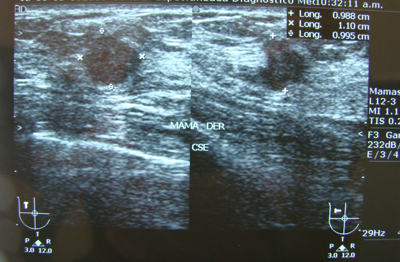
Ultrasound :1.1 cm lesion , irregular in one view and with augmented vascularity on Doppler .


Now the Radiology Diagnosis is a BIRADS IVa Lesion ( previously BIRADS II ) , enough to recommend a biopsy (Low probability) Yet her ObGyn performed a FNAB : 
ATYPICAL NEOPLASTIC CELLS.

Patient is refered to Oncology WITH a clear idea of being dealing with a BREAST CANCER.

Related Stress facing a BIRADS IV or V  is a COMMON CLINICAL SCENARIO in the Breast Clinic. Even some patients present an Important   Psycological Trauma. 

As a complement study : Digital Infrared Analysis was performed previous to Biopsy , with the following findings. 


Comparative Analysis of Both Breasts. Arrow: Clinically PALPABLE lesion , "invisible" for the INFRARED
Negative Predictive Value : PEACE OF MIND. 

Symetrical highest Fucntional Hyperthermias after the Cold Stress Test ( Cold Air) , Lesion at 9hrs of the right Breast : Absent or Invisible.

For some "negative" for the infrared analysis , for me a IR Score less than 100. 

Final statment in my practive : Although we have enough (Clinical , Radiological and from Citology )information for a complete removal of the lesion  we do know that your nodule is LOW in METABOLISM , LOW in AGGRESIVENESS even if positive for breast cancer , and MOST LIKELY : NEGATIVE FOR BREAST CANCER.(85% or more chances)

"So independently of the final results , you should be OK , you will be OK because the Odds are with you"


Comment : Final Pathology report : Simple Ductal Hyperplasia and a Benign Fibroadenoma. 

The Best ANATOMICAL image procedures in Breast are and probably will ever be : MAMMOGRAM(Rx) and ULTRASOUND  , the routine combination of both offers an adequate detection (when properly recommended).

Frequency and Initial Age for Screening are constantly being reviewed and are different for different Organizations and even differ between Countries. 

ANATOMICAL definition  depends on individual THRESHOLDS , and sometimes it is not clear enough in DIAGNOSIS , that is suspicion for Cancer : BIRADS IV(a,b or c) has the widest range of probability for breast cancer : 5-95% 

And even FNAB results : in this case Atypical Neoplastic , for some pathologist could represent POSITIVE for Breast Cancer .
And abscence of malignitiy in a FNAB does not rules out 100% Malignancy.
That is why some centers do not recommend it.

A PHYSIOLOGICAL  study like the Digital Infrared Analysis of the Breast offers aditional useful information of the clinically interest area.

Its NEGATIVE PREDICTIVE VALUE : Low score or "invisible" , non identified in the infrared specturm is strongly related to Abscence of Malignancy , or better yet  as a complementary study means in this case :

LOW or NULL  METABOLIC RATE


Clinically , a negative IR gives "irPeace of Mind" before Final Pathology Report ( In some centers , specially in developing countries this might take weeks)

"The DIRA of the Breast Shoul Be Routinely performed as a complementary Metabolic Procedure to Anatomical Standard Images and restricted to Certified Specialists in Breast Pathology : Oncologists , Radiologists , Breast Surgeons and Oncological ObGyn."

"Prospective controlled protocols would confirm this findings. "

EMC

Report of a case , high metabolic index . Microvascular density , lets help Mammography. Earlier Referal


63 year old Female , witha personal History more than 10 years ago of having an Stage II Infiltrating Ductal  Breast Carcinoma on her right side ,  treated with Radical Mastectomy plus standard adjuvant treatment with chemo and radiotherapy plus 5 years with oral tamoxifen.  
She presents  to oncological examination referred by a gyneocologist (November 2009)  , with a previous history of a nodule been detected by self breast examination in January 2009 and 10 months later she presents with the following clinical findings :  


Evident presence of a left breast nodule , not only palpable but visible  4-5 cm in diameter in the Upper External QUadrant of the remaining breast  . Clinically Positive Axillary Lymph Nodes around 1-2 cm  .


The nodule had  defined borders witha rubberish sensation at manipulation , obviously increased local temperature and even erythematous coloring of the skin .  

So initially , there is enough information to suspect either :  
  • Recurrent metastatic contralateral Breast Carcinoma  , with inflammatory component. OR:  
  • Primary remaining  Breast Carcinoma Cáncer de Mama Stage IIIB because of the inflammatory EC IIIB component. 


Yet ,  she presented with these previous radiological studies performed 9 months before :  

Analogic Mammogram of the remaning breast 


Complementary Ultrasound revealed 
 A solid , wider than taller  nodule , with regular borders . with out posterior enhancement or calcificacionts.  

Aproximately 2 cm in tis larger dimension. 

At DOPPLER : no considerable vascular flow .

Surprisingly 

INITIAL DIAGNOSIS , EVEN WITH THE PERSONAL HISTORY OF BREAST CANCER WAS : 

BIRADS II ,  WITH A BENIGN BREAST FYBROID  AS THE FIRST POSSIBILITY. 

Evidently , a radiological appreciation error or misdiagnosis 

YET , "MISTAKES TEND TO HAPPEN SINCE WE ARE ONLY HUMAN , OR NOT??" EMC.

Radiological Interpretation is a subjective Phenomena , with its concomitant corresponding misdiagnosis .

 l

10 months after , and after persistence and progression of the same exact lesion , the patient was referred with the following images:    


Evident radiological preogression , in size and density , yet with the same oval form. 


Complementary ultrasound now revealed a  4.6 cm nodule WIDER THAN TALLER 


Radiological Diagnosis Chaneged to : 
BIRADS V . 

As a complemetary procedure we performed DIGITAL INFRARED ANALYSIS  with the following images and metabolic implications.  


Infrared generation by the clinically suspected lesion is more than evident  its Thermal Summit reached  33.8 centigrades with a a DT to the Peripheral Tissue of more than  de 3 degrees  (3.3) and a  of a 191 points. 

So it is inferred a high metabolic index , an aggresive differentiation grade and or severe inflammatory component included and obviously the associated clinical consequences. 


Biopsy Results revealed: 

Angiosarcoma of the Breast. 


With Vascular Invasion.Comment: Angiosarcoma of the Breast is a rare malignant entity  , less than 1% of the total number of cancer cases . It is derived from mesenchymal tissue , specifically from blood vessels.It carries a poor prognosis since it has a fast doubling  time demostrated by NUMBER of mitosis or duplication rate. Metastases are common , and differing from Ductal Breast Carcinoma they are hematogenous not lymphatic.This sample case exemplifies , how if IR analysis would be offered as a complementary tool after Mammogram and Ultrasound it could have helped to a BETTER DETECTION , DIAGNOSE and EARLIER REFERAL (iReferal)to an Oncologist after Biopsy.INFRARED ANALYSIS IS NOT INTENDED TO SUSTITUTE XRAY OR MAMMOGRAPHIC EVALUATION , IS A PROCEDURE WITH A METABOLIC "MEANING" SPOKEN BY HIGHLY TRAINED SPECIALISTS AROUND MASTOLOGY.
"I believe it should be thoroughly reinvestigated and prospectively researched , in a multicenter study with an standarized procedure and comparative to standards of care procedures." "There is no harm doing it after Xrays or USG , on the contrary  : It could help or reaffirm and even offer different information given by detection or morphological studies. EMC"


miércoles, 23 de mayo de 2012

Case Number 7. Previous Lobullar Carcinoma. "Earlier Detection" , SyneIRgy" , "TABB"


 Case Number 7 . 


Female patient , Medical Dr: General Practicioner.  48 years old , with  familiar history in one aunt for breast cancer , 2 years previously  (2007)  during self exploration of an irregular right sided nodule she  attended to her physician  and after a difficult and irregular diagnosis algorithm  and a delay of 4 months , we decided to perform a biopsy :


 Invasive Lobullar Breast Carcinoma
 Negative for HER 2 neu and Hormone Receptors


Stage IIB :  T2N1M0 , she underwent a Radical Mastectomy , with a lesion of 4 cm and 2 positive lymph nodes and after wards standard adjuvant treatment


2009 after 2 years in control , conventional image studies were performed  :


Remaining Breast Mammogram  Lateral Oblique View  : dense breast tissue in her inferior quadrants  , superior isolated density with a "linear" aspect. 
 Cefalo Caudal view : persistance of the density in th upper and now external quadrant  . And internally an ovoidal  density
Close up for the superior density. No calcifications were seen. 


Magnification  of the upper and external density  , 
compatible with a linear " scar "image no 
calcifications were delineated either. .
Magnification of the internal lesion , opaque and ovoidal  


Ultrasound definition of the latter.  


Final Radiological Report  . 

BIRADS IV because of the presence of the INTERNAL LESION
Density towards the Spence tail of the breast
 was mentioned , yet specifically with out the 
need for a Biopsy. 

Digital Infrared Analysis as a complementary procedure : 


Evidence for an isolated Hiperthermia , that persists even after the cold sitmuli in the functional part of the study in the upper outer quadrant of the remaining breast (left) that is coincidental with the density defined in mammogram .


Suspicious by this method  with and IR Score of 125 , Peripheral Tissue delta between basal and functional studies of  1.3 degrees (obviously there is no  comparison with the contralateral breast )   


Close up for the IR  IMAGE , superior arrow corresponds to my radiological interesting area  and the reported by radiology  internal nodule (palpable) "invisible" or absent by this means. 


Which translates :  


  Suspicion for the upper lesion : Higher metabolic generation , vascular density , vasodilation , inflammation or infection . 


And  in the internal :  low heat or low  infrared radiation generation : low metabolic index  : low vascular density  , no inflammation or infection.

Having and informed consent ( patient is also a Medical Doctor)  we decided to perform both biopsies  . 


In the upper outer quadrant the IR area was delineated , lumpectomy 5 cm width was performed  down to the pectoralis major fascia . For it to be sure that no breast tissue was left behind.  


"Thermographically Assisted Breast Biopsy : TABB" you can google :" Biopsia de mama asistida por termografía

And complete biopsy of the internal nodule.


Macroscopically , the initial tissue of the lumpectomy was not revealing , yet the inferior was compatible  macroscopically and microscopically  with a benign breast fybroma.

Microscopical Images for the UPPER LUMPECTOMY  SPECIMEN: 


Definitive PAthological Report:

Atypical Lobullar Hyperplasia with , microcalcifications. Some other expert pathologist diagnosed it even as a lobullar carcinoma insitu. 


Comment: 
  • Lobullar Carcinoma is a neoplastic entity of difficult diagnosis , even with the most recent or advanced current image procedures. Its incidence is around 10% or less of the total number of cases.
  • So diagnostic mistakes are common  , even clinical palpable lesions are hard to define  with limits and consistency similar to an irregular "cushion"
  • Also it has a multiple lesion behaviour , in foci , centers or even bilaterally.
  • Hence metachronic lesiones are probable and common.
  • Atypical Lobullar Hyperplasia could progress  as high as 30% of the cases with an invasive form of the disease
  • Definitive treatment depends of common agreement and consensus with the patient and with the available resources , some recommend even PROPHYLACTIC  MASTECTOMY.

In this specific case  , she finally decided to have a Prophylactic treatment  so : Simple left mastectomy was performed and she started breast reconstruction.


By now she is well after 5 years of initial diagnosis and 2 years after completing breast reconstruction.  

Hypothesis:

Some Breast IR "promotors and supporters " sustain that Breast Thermography can detect lesions 8 to 10 years before Mammogram 

In my own point of view I believe this declaration is not believable and unrealistic and should be rejected by  Our scientific community (actually it is) . 


So we assume ,consider and agree that  this statement is  false doubtful  and even dangerous . 

"Yet not all of it is totally  incorrect ".EMC

Otherwise  , specific scenario exists :

  •  in high risk patients , 
  • with previous history of malignant lesions of difficult diagnostic and clinical behaviour  ,
  •  with doubt to recommend a  biopsy in the standard image studies 8mammogram and USG ) , 
  • Located in  most frequent site (statistically)for breast cancer : Upper External Quadrant. 


And coincidental between IR findings and mammographic ones: 

" DIGITAL INFRARED ANALYSIS OF THE BREAST IN EXPERT AND CERTIFIED HANDS, COULD HELP IN A SYNERGISTICAL WAY WITH THE CURRENT IMAGE PROCEDURES FOR THEM TO DETECT  EVEN  :

PREINVASIVE LESIONS" 

 EMC.

This specific case can only be seen by an experte in breast pathology , either an oncologist , breast surgeon o radiology trained a breast image expert.

So in this particular NICH of the breast cancer population  IR could  help  and  sustain therapeutical or eben prophylactical decisions

Finally

Mammography still is and will be the CORNER STONE of breast cancer detection through good quality screening program .


Yet :  Initial age of Screening  and it´s frequency is controversial even between experts  , it depends on race , cultural , economical and personal factors . 


It is also related to the corresponding  Breast Cancer Statistics for a given population , 


Human resources and quality of the available equipment , technique are also important  and finally the personal interpretation experience  is a decisive factor for it to be consistent-

Regretfuly , political and some enteprise interests are commonly involved. At least that is what I feel and find it not so hard to believe.


So:

"Mammogram :  Is a very complex medical diagnostic procedure."

Breast Thermography is a reproductible method , with objective values that could help synergistically  the available diagnostic armamentarium . 


Should be thoroughly re-studied  by Breast and Oncology Experts in  controlled , prospective protocols  and  preferably in mulcenter coordination. 

"Sine(IR)gy "EMC. 
"TABB ( Thermographically Assisted Breast Biopsy)

miércoles, 4 de abril de 2012

Case Number # 6. Young Patient High Risk Dense Tissue. Enhancing Breast Physical Exam

Case 6: 37 year old female , with family history 2 cousins and an aunt for breast cancer.
Three previous pregnancies , age of first birth at 30 , negative breastfeeding.
Contraception use for 1 year .
Asymptomatic until she claims discovering on self breast examination during shower a palpable nodule in the right upper quadrant.
At Physical Examination , a palpable , irregular , gritty lesion is detected in the right breast.
Axillary lumph node (1 ) palpable suspicious for malignancy.
After conventional studies (mammogram and ultrasound) she was sent to my office.

Mammogram:


Cefalo Caudal View : From this particular view it is impossible to define the clinicallly suspicious lesion.

CLOSE UP.
Medio Lateral Oblique View : Density assymetry in the upper right region close to the skin , without microcalcifications and undetermined axillary lymph nodes.

CLOSE UP, UNDETERMINED NOT WELL DEFINED DENSE LESION
.
Ultrasound definition : a 2.59 cm lesion , taller in it's larger dimension , irregular and solid suspicious for breast carcinoma classified as BIRADS IVc.

"Enough evidence to suspect malignancy and recommend immediate biopsy ".

Complemet Digital Infrared Analysis and findings :
CCan score of 173 for right breast .
With two functional persistent hyperthermias in the upper outer quadrant of the right breast , with an average of 32.7 in the interest area
Thermal summit of 33.6,
DELTA to surrounding tissue of 1.7 and 1.4 to the contralateral breast location

IT CAN BE ASSUMED EVEN BEFORE BIOPSY THAT THIS LESION CORRESPONDS TO A :

HIGH GRADE MALIGNANT LESION .


Frontal Basal Study View. Evident assymetry is observed at the Outer Upper Quadrant of the Right Breast , with augmented vascular network at this site.
Thermal or infrared SUMMIT DEFINED using isothermal standard option at software analysis.

Objective analysis of the interest area with results given in centigrades.
PINPOINT sign : Digital Infrared Analysis in this patient could easily direct the exact location of the palpable lesion.


Quadrantectomy and radical axillary dissection.
Close up : a gritty , white , irrgular 2.5 lesion is observed with negative margins .
FINAL PATHOLOGICAL REPORT:

- POORLY DIFFERENTIATED INVASIVE DUCTAL CARCINOMA , WITH SOME MEDULLARY CHARACTERISTICS.

- SIZE: 2.5.x2.2x2CM

- GRADE III IN MODIFIED SBR.

- WITH HIGH GRADE INVASIVE DUCTAL CARCINOMA I , SOLID TYPE WITH DUCTAL CARCINOMA IN SITU APROXIMATELY IN 20% OF THE NEOPLASM.

- WITH VASCULAR AND LYMPHATIC INVASION.

- NO NECROSIS .

- SKIN WITHOUT NEOPLASTIC CELLS.

- SOME FIBROCYSTIC CHANGES.


AXILLARY LYMPH NODES :

- 19 OUT OF 19 LYMPH NODES , NEGATIVE FOR NEOPLASTIC CELLS , WITH FOLLICULAR HYPERPLASIA ONLY .

PENDING IMMUNOHYSTOCHEMICAL RESULTS.

Comment:

40 years old or less patients and probably younger than 50 (depending on race and populations) , are condemned to FAILED early detection screening results based on mammogram because of physiologic DENSE TISSUE at this age , hence most of them will seek medical attention only after the lesion is clearly palpable either by self examination or by a trained health professional.

Even if the patient has a personal or familiar high risk background , this scenario usually repeats.

So current recommendations include earlier conventional studies ( 10 years or more earlier than the age for the Breast Cancer Relative (sister , mother , grandmother and second line ones , in this case cousins and aunt) .

HYPOTHESIS : IF DIGITAL INFRARED ANALYSIS OF THE BREAST DEPENDS ON METABOLIC , SIZE , AND PROBABLE DEPTH LOCATION OF THE NEOPLASTIC LESION .

AN ASSYMETRY SUCH AS THE ONE THIS CASE DEMONSTRATES COULD PROBABLE DETECT HIGH GRADE LESION AROUND 2 CM OR LESS : T1

YOUNG HIGH RISK PATIENTS YOUNGER THAN 40 (OR EVEN 50) COULD BE APPROACHED IN A MULTI-IMAGE MODALITY ( xRAY , ULTRASOUND AND DIRA).

DIRA COULD BE SPECIFICALLY RECOMMENDED IN THIS PATIENTS AS AN INTERVAL STUDY EVERY 6 MONTHS .

ANY EVIDENT INFRARED CHANGE COULD PROMPT FURTHER CONVENTIONAL STUDIES.

interval-hr-ir (High Risk -infrared)

AS SUCH , LESION COULD BE DETECTED NEAR A 1-2 CM IN SIZE , POSSIBLY HIGH GRADE

AND COULD BENEFIT THIS SPECIFIC POPULATION NICHE AND AT THE SAME TIME :

"ENHANCE BREAST EXAMINATION BY TRAINED HEALTH PERSONNEL."