Breast Digital Infrared Analysis (DIRA) , a forgotten and set aside procedure.

Visita: termografiamamaria.blogspot.com (Version en Español)

Breast DIRA (previously named Thermography) already proved in the past that it´s use as an screening procedure had low specificity and high sensitive values for it to be recommended as an standard care Breast diagnosis tool .
Yet , until recently (last 5 years) active research mainly by Surgical Oncologists or Breast Surgeons have published interesting results.( Cornell NY , Addenbrooke Cambridge , Ville Marie Montreal , Taiwan and Mexico CEPREC)
FDA previous published criteria were prohibiting DIRA as well as the A.C. of Radiologists ( as an initial SCREENING TOOL)

Yet it has been cleared by the FDA approval as an adjunctive screening tool for breast cancer .( under 510 k) .

DIRA´s meaning is not an ANATOMIC or ARCHITECTURAL procedure ,it is a METABOLIC or PHYSIOLOGIC ONE so at first it ´s current recommendation should be only as a COMPLEMENTARY STUDY.

As such DIRA could help in specific scenarios, BIRADS 0,3,4 and give potential useful additional data in BIRADS5. EVEN BEFORE BIOPSY
Could aid mammogram in young,dense and breast prosthesis patients, and should be evaluated in other clinical scenarios as a Neoadyuvant Monitoring tool.
Finally as an early detecion procedure specifically in underdeveloped countries , could Triage in search of aggresive forms of the disease(T1,G2_G3). And aid Physical Exam of the Breast .
This site is dedicated to open mind Breast specialists ONLY .
¨EXPANDING OUR CURRENT COMPREHENSION, UNDERSTANDING AND APPROACH OF IMAGE AND DIAGNOSIS IN BREAST CANCER¨

miércoles, 30 de enero de 2013

Angiogenesis Related to Breast Cancer , could it be Measured?

One of the most frequent arguments Infrared Supporters mention throughout the web is that Angiogenesis that appears in Breast Cancer is able to rise local temperature so that  the Infrared Detector could actually see and Measure.........

Is this principle correct? 

Is angiogenesis capable of an abnormality enough for it to be seen and recorded?

Oncological Physiology Mentions  that Angiogenesis is a determinant factor for some tumors to hold on and then grow. 

It seems quite logical , it has been proven and as such we must accept......

Yet Breast Cancer is an Heterogenous Disease , meaning that there are different types of Breast Cancers and that some may not have Neoangiogenic Factors or Enough Microvascular Density. 

As a matter of fact the latter is just a weak prognostic factor in breast cancer.... so I believe that Angiogenesis is NOT THE ONLY FACTOR that rises Infrared Radiation from a given Tumor or Cancer.

Nevertheless I will present a clinical finding that supports the principle of angiogenesis and Infrared Detection........

Sample Case :

Female patient in her early 40´s, previous history of RADIATION Therapy in her Right Thigh during childhood for a Liposarcoma  more than 25 years later Metachronic ANGIOSARCOMA , Treated in the US , with chemotherapy , surgery and TRAM FLAP  plus radiation , yet clinical evidence for Recurrence a year after being in control .

Infrared Analysis just to have an simple image:


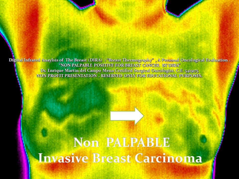
Infrared Analysis of the Anatomical Region. Clear Differences in Color are seen as Infrared Radiation Rises. Advanced and current State of the art IR detectors have a LARGER color scale, resolution and pixels.


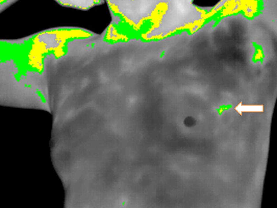
Software Color and Palette options can can choose only gray scale , white and black:

 And with the Use of Automatic "ISTOTHERMS" or color ranges , the highest IR radiation areas are limited and subsecuently could be measured.

Software manipulation and applications can give objective numerical reproductible results and hence record them and compare if future instances occur.

Angiogenesis in Angiosarcoma is EVIDENT , local heat is risen because of VASCULAR DENSITY and in part I assume could explain what happens in SOME TYPES of Breast Cancer.

If this should be enough to be present at Breast´s Skin  Surface and be a CLEAR sign that actually means malignancy is for Prospective Controled Clinical Protocols to be defined.

IR Analysis is related to metabolic , inflammatory , infectious and vascular biological conditions of the focused area.......

These images Mean NOTHING to Most Common clinicians and or Oncologists , they do not change their treatment algorithm or way to make a diagnosis , are useless , time consuming and considered fraudulent........

Are they really?

But they proof at least ONE Physiological and Pathological principle : Angiogenesis.

Time to think..........

miércoles, 16 de enero de 2013

Ductal Carcinoma In situ , Clearly Visible by Digital Infrared Analysis (IRDCIs)

Good Evening , Is Ductal Carcinoma In Situ   of The Breast Visible under the infrared Spectrum ?
Just authorized by the patient to publish her images?Ready?

40 Year Old Female , Never Pregnant , no Family History for Breast Cancer. 

Now we will start backwards:
Digital Infrared Analysis First ok?


Basal Anterior View or Frontal . Total Breast areas are included. Assymetry is focused mainly towards The Left Breast Upper and Outer Quadrant.

Basal ( Previous to Cold Challenge) Right Oblique View  and Left Oblique View
Arrow points toward the "highest" Infrared Radiation Area. Difficult to see and detect for the "untrained" eye. 


Physiologic or Functional (After Cold Stress Test or Challenge ) . Now a Clearly Defined area is EASILY detected (Arrow) and correponds to the Highest Metabolical Active Area of BOTH BREASTS. 

Would You Lke to See the Ultrasound and Xray Images?
Keep Posted......
Here are the Ultrasound Images:
 IRRELEVANT ULTRASOUND IMAGES , SIMPLE CYST AT MOST
Now here comes our Cornerstone in Detection :
Left superior and External SUSPICIOUS MICROCALCIFICATIONS.
With slightly dense surrounding tissue. 
BIRADS IV you all will agree. (I hope)


NOW REMEBER THE INFRARED SERIES PREVIOUS TO BIOPSY:

Go back to see larger details.......

Finally patient was Biopsied under Radiological Guide :
Mammography strength also rely in the ability to locate EXACTLY the anatomic position of the detected "abnormality".....
YET , although it may seem improbable or unbelievable , this latter kind of radiological maneuver is NOT present or available in MOST countries.
All efforts are stablished to OFFER radiological diagnosis and assitance for surgical or biopsy procedures. 
BUT Real clinical environments , and surrounding complex cultural and economical parameters make XRay detection and diagnosis of breast cancer highly unlikely to occur.

But then again WHO cares for women with no access to mammography or those who do not have access to a good radiologist or cannot AFFORD a guided Biopsy or even a simple Biopsy?


MMmmmmmm. Makes me wonder..........

But , back to our patient.
Final Pathological Report:

Ductal Carcinoma In Situ.

Patient chosed a Quadrantectomy which was reconstructed with autologous Latissimus Dorsi Myocutaneous Flap.
 Plus adjuvant therapy and now she is having Tamoxifen 20 mg a day.

2 Years Later , undercontrol She is doing fine and well as the vast majority of Ductal Carcinoma in Situ patients are.

Comment: Some In Situ Lesions might be clearly visible under the infrared spectrum (IRDCis), prospective controlled clinical trials could define which early lesions are clearly defined.

It is not to fight Mammography , it has never been the issue. IR analysis reintroduced in expert trained Breast Specialist could help against Breast Cancer.

I know what you might say or think , there is nothing done different from what is usually done. 

You are absolutely right , IR image could be thought as USELESS or TIME consuming etc....

Yet it has a meaning at could actually HELP.

One final question :

 My patient is doing well BECAUSE Mammography Detected an Early Breast Cancer Stage 0?
Or
Is she doing fine BECAUSE the Kind of Cancer That we found was never going to do any harm ?

pffffffff : I do not Know , I guess somewhere in Between.....

Can we Reduce OVERDIAGNOSIS and OVERTREATMENT?

Have a great day.........


History and some Facts to Think About

Yesterday  , I was Surfing Through the Internet As we all do .........searching the topics we like the most.

It suddenly reached my attention the Following In Memoriam:


Harold J. Isard MD.

Pioneer in Roentgenology , Early Breast Cancer Detection and Curiously an active Researcher of Thermography (1970,s) with:

 A VERY  INTERESTING DISCUSSION & SUMMARY.

BUT FIRST A PERSONAL COMMENT , JUST TO MAKE IT CLEAR:


"I support the use of Mammography as the cornestone procedure for breast cancer morphological detection , its frecuency and age to do it vary greatly and depend on multiple variables and environments. 

It is indicated most certainly individualy , depending mostly on risk factors and local statistics. 

Finally clean , ethical  , expert clinical judgement after useful information after image procedures render the better understanding , certified recommendations and possible outcome. 

Thermography stands alone as a potential metabolical , vascular , inflammatory or infectious study that can help against breast cancer" EMC

Now Dr. Isard mentions :
  1. He affirms that THERMOGRAPHY Cannot diagnose cancer , but hey Only Pathology diagnoses Cancer.
  2. He states that THERMOGRAPHY can obtain ABNORMALITIES......
  3. He sustains even then that MORTALITY HAS NOT CHANGED ( And guess What , according to NEJM latest Review it Seems that AFTER 30 years of Screening with Mammography MORTALITY HAS NOT CHANGED THAT MUCH EITHER) Hard to believe but the facts have been published recently.....Controversy goes on 
  4. He suggests Thermography as a PRELIMINAR STUDY before Physical Examination and Then Mammogram ( SOUNDS REALLY CRAZY I MUST RECOGNIZE  and OUR DETECTION and DIAGNOSTIC APPROACH ALGORITHM has changed quite a bit since then.
  5. No wonder WHY THERMOGRAPHY was put aside , IMAGES were REALLY  CONFUSING , digital era may give it a new opportunity.
  6. He mentions Thermographic difficulties for specificity , REMEMBER BIRADS IV is 5-95% specific as well.
  7. He Highlights Vascular Patterns.
  8. He Remarks how CONSTANT and UNCHANGED the thermal PATTERN is.
  9. Regardless of the Date During The Menstrual Cycle , the "patient" or volunteer could be EASILY RECOGNIZED and Hence abnormalities also could be defined.... and monitored.
  10. Guess what : he mentions that the Thermal Image is RELATED to the BIOLOGICAL BEHAVIOUR of the SUSPECTED LESION........I constantly repeat this statement through my blog
  11. He identified IN SITU  lesions as well as Metastatic. I will show you some of this later .....future cases.
  12. "The DEVELOPMENT of an ABNORMAL PATTERN  when compared to a normal baseline study must ALWAYS be viewed with extreme suspicion" I have a sample case....
  13. Emphasizes Clinical JUDGEMENT to reduce UNNECESARY SURGERIES ( of course we now have minimal invasive procedures that can be applied) yet interesting point.
  14. Thermography was DIVIDED into POSITIVE or NEGATIVE ONLY , I believe its meaning is METABOLIC then its contribution has to be METABOLIC ALSO . 0 or 1 seems too simple.
  15. Thermography ALONE Cannot Beat MAMMOGRAPHY.......That is very clear for me and for everyone no controversy on that.....(specificaly on morphological meaning.....)
  16. Yet he mentions :"Since Thermography  and Mammography did not always suspect the SAME lesion , the USE OF BOTH studies INCREASED ACCURACY to 92% when either or both examinations were POSITIVE.How about that....
  17. Thermography ACTUALLY IMPROVED Clinical Examination, now if we still Recommend Clinical Examination CAN WE REINTRODUCE Thermography during the Highly Specialized Breast Physical EXAM????
  18. BACK then CONTRALATERAL CLINICALLY NEGATIVE BIOPSIES were Performed , astonishingly some of them Positive for Beast Cancer...........so CANCERS EXIST but they do no harm EVER? Back to NEJM recent DATA.}
  19. With Thermology Technology in Those days  : 61% of asymptomatic patients with cancer were identified , I wonder if now Digital Infrared Analysis of the Breast Can do Better?
  20. Thermography Improves DIAGNOSTIC ACCURACY , for me only used by and reserved for  ONCOLOGISTS.
  21. Could EMERGE as a preliminary Screening Procedure .....Uuuffffff.....SHAKY GROUNDS indeed : Heavy evidence against this proposal , greater economical interests and maybe Political as well. Not to mention Status Quo and Orthodox Mentalities....
  22. Mammography detects around 1-6 cases per 1000 of screened women , I believe this ratio is still the same . But if I am wrong please do not hesitate to correct me.
  23. Mammography PLUS Thermography 7.3-1000 ( Can THIS SINERGY be Better  Nowadays?)
  24. Here comes the TRICKY NUMBERS if USED as a PRELIMINARY STUDY that is BEFORE MAMMOGRAPHY:   21 cases per 1000 mammograms. This would mean BETWEEN 3 to 20 Times FEWER mammograms done. (uupppssss    oh oh  do the NUMBERS please)
  25. BUT  : 10 cancers were NOT DETECTED By IR , and that is the reason for NON STOP MASSIVE MAMMOGRAPHIC XRAY SCREENING , I think these 10 cancers would have a BETTER prognosis even if undetected , I suspect LOW METABOLIC HEAT and a "Benign" Biological Behaviour........sounds reasonable? In other words maybe those not identified do not actually kill anyone.......interesting.
  26.  And finally he states the INHERENT difficulties for an OPTIMAL MAMMOGRAPHY SCREENING PROGRAM.


 In my real world (and 95% of the world "screenable"population ) Optimal Screening is far from standard recommendations , coverage is practicaly ZERO , and we are loosing the battle as epidemiologists state , tumor size is around 5cm or more and detection campaigns throw breast cancer patients into the "system" and regretfuly they receive attention 6 months later , at best.

Reality Bites.......

Can we focus attention in Female Patients with Suspicious PATTERNS?

Can we Define this PATTERNS AGAIN PROSPECTIVELY? With the help of Mammography? 

After all the common enemy is BREAST CANCER , right?

I am not a professional editor , I will try to do my Best......

Hope you like it and probably some one Open Minded  Enough could be Professionally and Prospectively Re-interested.

BOY!!!!!!  

 I MISS THIS KIND OF RADIOLOGISTS , Do you happen to know ONE?


Hope you enjoyed it as much as I did......


lunes, 14 de enero de 2013

Multiple Bilateral Macrocalcification Associated with a Nodule.


Every once in a While under Mammogram plus Ultrasound Screening we can find Bilateral 

Macrocalcifications and Cysts that can confuse even the most expert Radiologist. Or a Radiologist in a Hurry might miss it.

Yet patients arrive to the Breast Clinic with the Following Images:

 Dense Breasts , multiple macrocalcifications  Bilateral Nodules
Inflammatory Bilateral Axillary Lymph Nodes  
12 hr Posterior Dense Assimetry of the Right Breast  , with Macrocalcifications.

 My Diagnosis  without Ultrasound : BIRADS 0

WOULD YOU LIKE TO SEE THE INFRARED IMAGE BEFORE ULTRASOUND ?


The Infrared Analysis before Ultrasound offer from my own point of view: Importante Metabolic , vascular , neoplastic or inflammatory information.

In this specific scenario , when the image is "moving" or has "moved" to one side CLEARLY  , the Ultrasound Exam and clinical focus follows it.

" THE INFRARED IMAGE COULD ASSIST ULTRASOUND, SEARCHING HIGHLY METABOLIC LESIONS. " EMC

"I believe coupling this two technologies can offer a better advantage that each one of them separately" EMC
 Clear Assimetry found on the Basal Anterior View.
Rectified Right Mammary Fold  and a Subtle Indentation (Arrow)

Basal Left Oblique View , Clear Lesser Vascular Network.
Physiological , Functional or Cold Stress Test View: Subtle Assimetry between Folds (Green Line on Isotherms)
Unique Persisten Unilateral Hyperthermia on the Functional Series.
Rectified Inferior Right Mammary Fold (Arrow) : Resembles the sign that can also be seen on Mammograms and translates Parenchymal Retraction from Tumor Invasion to Cooper Ligaments. 

Would you like to see the Ultrasound?

Right Breast  Line B-C  6hrs Solid Lesion , Wider than Taller around 1.8cm in its longesta diameter. With Doppler: 
Little if non Peripheral Vascular Flow  Enhancement. 
Irregular profile , seems almost circular in this view.

Although It might seem a possible Fibroadenoma , for most Breast Specialist is Definetly Suspicious enough for maybe lets say a BIRADS IVa , SO DETECTION WOULD BE COMPLETE , YET DOUBT ABOUT HISTOLOGY WILL RISE.

With previous Infrared Images and BEFORE biopsy clinical suspicion is ALSO HIGHER. 

My Clinical Comment was : "Mrs. X , there is definetly something going on in your right breast , FOR ME it is highly likely that it will be a Breast Carcinoma Indeed , possibly associated to a previous Fibroadenoma (which is very rare) , yet Lymph Nodes on palpation and in Xray are likely to be safe or negative for Cancer."

"We better get prepared , Histology confirmation is a must , the sooner the Better."

I know what you might think , if conventional studies were done detection would also be possible. 

You are absolutely right , you might even consider IR image
  • Obsolete
  • Meaningless
  • Waste of Money
  • Time Consuming , 
  • Out of Evidence , 
  • Done by Quacks , 
  • Not paid by Medical Insurance and  
  • Probably subject to liability.


Again you maybe right , but I Think IR image:
  1. Holds a Meaning , unknown to almost every certified breast specialist
  2. It Can Help Guide secondary image procedures.
  3. Talks About Metabolic Activity around or within the detected lesion
  4. In expert hands and done Ethically can stablish and enhance Empathy between Dr and Patient 
  5. Has Predictive Values that are interesting enough to be reinvestigated prospectively.
  6. It is Fascinating and for me  opens a new horizon in Breast Cancer Image. 
And for these reasons we should "talk" or "start to talk" Infrared Language. 

Finally , correct me if I am wrong: 
  • Radiologists FOCUS on DETECTION , they should.
  • Clinical Breast Specialists (vg. ObGyn´s) and Pathologists FOCUS on DIAGNOSIS , they should.
  • We Oncologists FOCUS on TREATMENT , we should.
"I believe IR can be applied in each of the previous 3 statements"EMC

Good Day.


miércoles, 9 de enero de 2013

Breast Thermography / Digital Infrared Analysis ,Historical and Personal Review.

The following is a Personal and Historical Review SLIDE SHARE presentation exact copy of the Webinar Held recently in NYC. I was Kindly invited by PACT ( the Professional Academy of Clinical Thermology ) to talk about my Digital Infrared Analysis of the Breast or Thermography as a Surgical Oncologist.

http://www.slideshare.net/drmartindelcampo/breast-thermography-digital-infrared-analysis-of-the-breast

Intented to the "other" Healthcare providers that currently support the use of Breast Thermography at first , but open and meant also to "our" certified oncological experts.

As I mentioned to the Director of PACT , if Infrared Image moves Forward in Breast Pathology , that would only mean that current users will not be able to perform it.

After all , Breast Cancer Care Responsability Relies on "US"

  • Oncologists
  • Radiologists
  • OBGyn´s
  • Breast Surgeons
  • Chemotherapists and 
  • Radiotherapists
It should be critised and rejected , but I think that it also deserves a chance for Controlled Clinical Trials.

It has never been meant to stand AGAINST mammography , after all even Ultrasound , Nuclear Medicine images and  MRI strengths  rely  in on it.

"Mammography is our cornerstone breast image procedure. Everything else should be built after it"

irImage is an Unknown Language with and unknown meaning , historically divorced from our Diagnostic Armamentarium. Yet recently : costs , sensitivity and digital performance have been improved.

We should research thoroughly and prospectively.......

http://www.slideshare.net/drmartindelcampo/breast-thermography-digital-infrared-analysis-of-the-breast

Hope you like it..........

lunes, 7 de enero de 2013

2011 National Oncology Congress Slide Show

http://www.slideshare.net/gusgarcia505/presentacin-smeo


For you , for free , though it is is in Spanish it is almost self explaining.

It does not represents standards of care in Mexico.

It should be used only for educational purposes only , and for possible future multicentric prospective controlled trials in certified breast or oncological hands. 

EMC

http://www.slideshare.net/gusgarcia505/presentacin-smeo

miércoles, 2 de enero de 2013

Non Palpable Breast Cancer Detected also by IR


BEST WISHES FOR 2013.

We Oncologists , ObGyns , Radiologists , Breast Specialists and Generally all Women and Population Believe and KNOW that the SMALLER the Tumor is The BETTER the prognosis.

Anatomical definition of Mammography sporadically (in my experience) detects invasive  tumors less than 1 cm.But I am guessing , correct me if I am wrong , that most international breast specialists have little experience in SMALL tumors.

So , prognosis is related directly to small size and viceversa..

Mammography STRENGTH around 50 to 69 years is such that would be useless to recommend anything else , or even try to atttack its controversies  , flaws , overdiagnosis , pain or possible radiation damage.

I support firmly Mammogram for detection in this specific group .

YET AROUND HALF THE BREAST CANCER POPULATION IS 50 YEARS OR LESS.

 In this setting STATISTICS and BENEFITS of Screening are in doubt or have not changed at all after 30 years (40y and less) , mainly because BREAST DENSITY

Optimal Screening Coverage in developing countries differ enormously it ranges below 10% when we know that the ideal percentage is around 70 and a minimum of 10 years of continuos screening .

Even more , its recommended Frequency is in review and different recommendations occur internationally.

I believe THE COMMON ENEMY IS CANCER , to add a possible tool against it means no harm and should not be rejected. The NEW benefit of the doubt should overcome . In order to learn its meaning and possible applications it is a TASK meant  for ALL.

So lets begin 2013 with this specific scenario : NON PALPABLE LESIONS that are visually detected by the DIGITAL INFRARED ANALYSIS.

By  NO MEANS i recommend (for now) as a substitute for a Mammography  , this images images represent a small piece of my Universe , only if a Mulcentric research in developed countries would be involved this piece of evidence could be reproduced.

They are MEANT TO BE the cornerstone for future research.

What should we do between Mammograms (2 or 3 year) changing intervals?

You should be the judge.


I Know it sounds RIDICULOUS or even ABSURD , believe me I started with the same doubts and rejection. After all I was not even told during my Oncological Training that this could be possible.

Yet this images are the result of a Blinded Challenge in order to Stablish if I could recognize the presence or abscence of the detected lesion ( ALREADY BIASED) I should mention.

Yet ,  for me it is Very Clear , Assymetry , vascular network and persistent Hyperthermias after the cold Challenge , plus axillary temperature and possible mammary fold characteristics
 (EMC Infrared Pentad for Breast Cancer ) 

REPRESENT A HIDDEN CLUE OR LANGUAGE FOR INVASIVE BREAST CARCINOMA.

Obviously THIS IMAGES HAVE TO BE be redefined and reconfirmed .

But : 

IF THEY ARE CONSTANT for 1-2 cm NON PALPABLE INVASIVE BREAST CANCER.Specially Aggresive forms : Grade 2 , 3 or HER2neu+++.

My Hypothesis is that DIRA could actually Help Detection , Downstage Breast Cancer Statistics and most of all Help Women.

Digital Infrared Analysis of the Breast SHOULD be reexplored  and resitricted only to Certified Breast Specialists. 

Fortunately there is only one option to confirm if what I was able to Find is true:  that would be forward prospective controlled trials to define non palpable breast Cancer under the infrared spectrum and analysis  : emartindelcampo@yahoo.com

Have a great 2013..