Every once in a While under Mammogram plus Ultrasound Screening we can find Bilateral
Macrocalcifications and Cysts that can confuse even the most expert Radiologist. Or a Radiologist in a Hurry might miss it.
Yet patients arrive to the Breast Clinic with the Following Images:
.JPG)
Dense Breasts , multiple macrocalcifications Bilateral Nodules
.JPG)
Inflammatory Bilateral Axillary Lymph Nodes
.JPG)
12 hr Posterior Dense Assimetry of the Right Breast , with Macrocalcifications.
My Diagnosis without Ultrasound : BIRADS 0
WOULD YOU LIKE TO SEE THE INFRARED IMAGE BEFORE ULTRASOUND ?
The Infrared Analysis before Ultrasound offer from my own point of view: Importante Metabolic , vascular , neoplastic or inflammatory information.
In this specific scenario , when the image is "moving" or has "moved" to one side CLEARLY , the Ultrasound Exam and clinical focus follows it.
" THE INFRARED IMAGE COULD ASSIST ULTRASOUND, SEARCHING HIGHLY METABOLIC LESIONS. " EMC
"I believe coupling this two technologies can offer a better advantage that each one of them separately" EMC
 Clear Assimetry found on the Basal Anterior View.
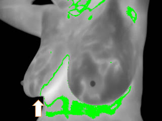
 Rectified Right Mammary Fold and a Subtle Indentation (Arrow)


 Right Breast Line B-C 6hrs Solid Lesion , Wider than Taller around 1.8cm in its longesta diameter. With Doppler:

The Infrared Analysis before Ultrasound offer from my own point of view: Importante Metabolic , vascular , neoplastic or inflammatory information.
In this specific scenario , when the image is "moving" or has "moved" to one side CLEARLY , the Ultrasound Exam and clinical focus follows it.
" THE INFRARED IMAGE COULD ASSIST ULTRASOUND, SEARCHING HIGHLY METABOLIC LESIONS. " EMC
"I believe coupling this two technologies can offer a better advantage that each one of them separately" EMC


Basal Left Oblique View , Clear Lesser Vascular Network.

Physiological , Functional or Cold Stress Test View: Subtle Assimetry between Folds (Green Line on Isotherms)

Unique Persisten Unilateral Hyperthermia on the Functional Series.

Rectified Inferior Right Mammary Fold (Arrow) : Resembles the sign that can also be seen on Mammograms and translates Parenchymal Retraction from Tumor Invasion to Cooper Ligaments.
Would you like to see the Ultrasound?


Little if non Peripheral Vascular Flow Enhancement.
Irregular profile , seems almost circular in this view.
Although It might seem a possible Fibroadenoma , for most Breast Specialist is Definetly Suspicious enough for maybe lets say a BIRADS IVa , SO DETECTION WOULD BE COMPLETE , YET DOUBT ABOUT HISTOLOGY WILL RISE.
With previous Infrared Images and BEFORE biopsy clinical suspicion is ALSO HIGHER.
My Clinical Comment was : "Mrs. X , there is definetly something going on in your right breast , FOR ME it is highly likely that it will be a Breast Carcinoma Indeed , possibly associated to a previous Fibroadenoma (which is very rare) , yet Lymph Nodes on palpation and in Xray are likely to be safe or negative for Cancer."
"We better get prepared , Histology confirmation is a must , the sooner the Better."
I know what you might think , if conventional studies were done detection would also be possible.
You are absolutely right , you might even consider IR image
- Obsolete ,
- Meaningless
- Waste of Money
- Time Consuming ,
- Out of Evidence ,
- Done by Quacks ,
- Not paid by Medical Insurance and
- Probably subject to liability.
Again you maybe right , but I Think IR image:
- Holds a Meaning , unknown to almost every certified breast specialist
- It Can Help Guide secondary image procedures.
- Talks About Metabolic Activity around or within the detected lesion
- In expert hands and done Ethically can stablish and enhance Empathy between Dr and Patient
- Has Predictive Values that are interesting enough to be reinvestigated prospectively.
- It is Fascinating and for me opens a new horizon in Breast Cancer Image.
And for these reasons we should "talk" or "start to talk" Infrared Language.
Finally , correct me if I am wrong:
- Radiologists FOCUS on DETECTION , they should.
- Clinical Breast Specialists (vg. ObGyn´s) and Pathologists FOCUS on DIAGNOSIS , they should.
- We Oncologists FOCUS on TREATMENT , we should.
"I believe IR can be applied in each of the previous 3 statements"EMC
Good Day.
No hay comentarios:
Publicar un comentario