Case Number 7 .
Female patient , Medical Dr: General Practicioner. 48 years old , with familiar history in one aunt for breast cancer , 2 years previously (2007) during self exploration of an irregular right sided nodule she attended to her physician and after a difficult and irregular diagnosis algorithm and a delay of 4 months , we decided to perform a biopsy :
Invasive Lobullar Breast Carcinoma
Negative for HER 2 neu and Hormone Receptors
Stage IIB : T2N1M0 , she underwent a Radical Mastectomy , with a lesion of 4 cm and 2 positive lymph nodes and after wards standard adjuvant treatment.
2009 after 2 years in control , conventional image studies were performed :
 |
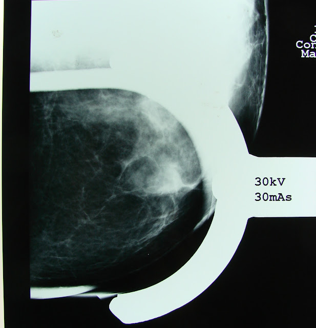
Remaining Breast Mammogram Lateral Oblique View : dense breast tissue in her inferior quadrants , superior isolated density with a "linear" aspect. |
Cefalo Caudal view : persistance of the density in th upper and now external quadrant . And internally an ovoidal density
 |
| Close up for the superior density. No calcifications were seen. |
 |
Magnification of the upper and external density ,
compatible with a linear " scar "image no
calcifications were delineated either. .
|
 Magnification of the internal lesion , opaque and ovoidal
 |
| Ultrasound definition of the latter. |
 |
Final Radiological Report .
BIRADS IV because of the presence of the INTERNAL LESION
Density towards the Spence tail of the breast
was mentioned , yet specifically with out the
need for a Biopsy.
Digital Infrared Analysis as a complementary procedure :
|

Evidence for an isolated Hiperthermia , that persists even after the cold sitmuli in the functional part of the study in the upper outer quadrant of the remaining breast (left) that is coincidental with the density defined in mammogram .
Suspicious by this method with and IR Score of 125 , Peripheral Tissue delta between basal and functional studies of 1.3 degrees (obviously there is no comparison with the contralateral breast )

Close up for the IR IMAGE , superior arrow corresponds to my radiological interesting area and the reported by radiology internal nodule (palpable) "invisible" or absent by this means.
Which translates :
Suspicion for the upper lesion : Higher metabolic generation , vascular density , vasodilation , inflammation or infection .
And in the internal : low heat or low infrared radiation generation : low metabolic index : low vascular density , no inflammation or infection.
Having and informed consent ( patient is also a Medical Doctor) we decided to perform both biopsies .
In the upper outer quadrant the IR area was delineated , lumpectomy 5 cm width was performed down to the pectoralis major fascia . For it to be sure that no breast tissue was left behind.
"Thermographically Assisted Breast Biopsy : TABB" you can google :" Biopsia de mama asistida por termografía
And complete biopsy of the internal nodule.
 Macroscopically , the initial tissue of the lumpectomy was not revealing , yet the inferior was compatible macroscopically and microscopically with a benign breast fybroma.
Microscopical Images for the UPPER LUMPECTOMY SPECIMEN:
.JPG)
.JPG)
.JPG) |
Definitive PAthological Report:
Atypical Lobullar Hyperplasia with , microcalcifications. Some other expert pathologist diagnosed it even as a lobullar carcinoma insitu.
Comment:
- Lobullar Carcinoma is a neoplastic entity of difficult diagnosis , even with the most recent or advanced current image procedures. Its incidence is around 10% or less of the total number of cases.
- So diagnostic mistakes are common , even clinical palpable lesions are hard to define with limits and consistency similar to an irregular "cushion"
- Also it has a multiple lesion behaviour , in foci , centers or even bilaterally.
- Hence metachronic lesiones are probable and common.
- Atypical Lobullar Hyperplasia could progress as high as 30% of the cases with an invasive form of the disease
- Definitive treatment depends of common agreement and consensus with the patient and with the available resources , some recommend even PROPHYLACTIC MASTECTOMY.
In this specific case , she finally decided to have a Prophylactic treatment so : Simple left mastectomy was performed and she started breast reconstruction.
By now she is well after 5 years of initial diagnosis and 2 years after completing breast reconstruction.
Hypothesis:
Some Breast IR "promotors and supporters " sustain that Breast Thermography can detect lesions 8 to 10 years before Mammogram
In my own point of view I believe this declaration is not believable and unrealistic and should be rejected by Our scientific community (actually it is) .
So we assume ,consider and agree that this statement is false , doubtful and even dangerous .
"Yet not all of it is totally incorrect ".EMC
Otherwise , specific scenario exists :
- in high risk patients ,
- with previous history of malignant lesions of difficult diagnostic and clinical behaviour ,
- with doubt to recommend a biopsy in the standard image studies 8mammogram and USG ) ,
- Located in most frequent site (statistically)for breast cancer : Upper External Quadrant.
And coincidental between IR findings and mammographic ones:
" DIGITAL INFRARED ANALYSIS OF THE BREAST IN EXPERT AND CERTIFIED HANDS, COULD HELP IN A SYNERGISTICAL WAY WITH THE CURRENT IMAGE PROCEDURES FOR THEM TO DETECT EVEN :
PREINVASIVE LESIONS"
EMC.
This specific case can only be seen by an experte in breast pathology , either an oncologist , breast surgeon o radiology trained a breast image expert.
So in this particular NICH of the breast cancer population IR could help and sustain therapeutical or eben prophylactical decisions
Finally :
Mammography still is and will be the CORNER STONE of breast cancer detection through good quality screening program .
Yet : Initial age of Screening and it´s frequency is controversial even between experts , it depends on race , cultural , economical and personal factors .
It is also related to the corresponding Breast Cancer Statistics for a given population ,
Human resources and quality of the available equipment , technique are also important and finally the personal interpretation experience is a decisive factor for it to be consistent-
Regretfuly , political and some enteprise interests are commonly involved. At least that is what I feel and find it not so hard to believe.
So:
"Mammogram : Is a very complex medical diagnostic procedure."
Breast Thermography is a reproductible method , with objective values that could help synergistically the available diagnostic armamentarium .
Should be thoroughly re-studied by Breast and Oncology Experts in controlled , prospective protocols and preferably in mulcenter coordination.
"Sine(IR)gy "EMC.
"TABB ( Thermographically Assisted Breast Biopsy)
|


 Magnification of the internal lesion , opaque and ovoidal


.JPG)
.JPG)
.JPG)