Breast Thermography CANNOT DETECT Clear Mammographical Microscopic Breast Carcinomas , yet (Biological information as a complement is important in the Doctor-Patient Relationship. And offers predictive biological behaviour of the lesion prior to biopsy , useful for both.) EMC
Would you like to see an Example??
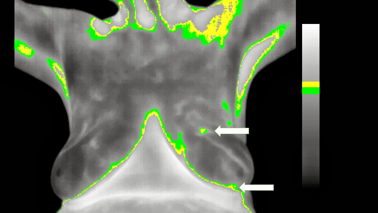
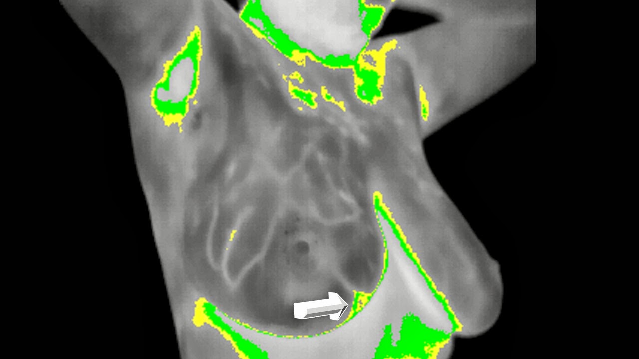
Early 40´s asymptomatic woman , first mammography screening.
On Physical Examination after the images an irregular "soft" lump was felt on her right inner upper quadrant......
Anatomically Clearer than this , is Impossible and that is why detection is supported on MORPHOLOGY.
Makes more than sense....
So would you perform an Infrared Complementary Procedure???
Vast Majority : will say ABSOLUTELY NOT....Right?
Well I did , why :
BECAUSE EVEN BEFORE BIOPSY I HAVE USEFUL INFORMATION FOR THE PATIENT TO GIVE.......
METABOLIC INFORMATION....Makes sense to me .....( and to tell you the truth it is brainstimulating......)
"EMPATHY , PREDICTION , COMPLIANCE , STRESS , AND IN SOME CASES EVEN THERAPEUTIC DECISIONS CAN BE SUPPORTED BY AND INFRARED IMAGE."
EMC.
I believe you should learn it TOO.... don´t you think so ???
To see the IR images Keep posted OK??? , have a great week......
Non profit site dedicated to the active research and open share of our INFRARED Oncological results and experience. IF YOU LIKE THE INFORMATION PLEASE SHARE AND FORWARD IT...... ¨Reintroducing Infrared Image , to Aid our Current Understanding of image in Breast Cancer¨
Breast Digital Infrared Analysis (DIRA) , a forgotten and set aside procedure.
Visita: termografiamamaria.blogspot.com (Version en Español)
Breast DIRA (previously named Thermography) already proved in the past that it´s use as an screening procedure had low specificity and high sensitive values for it to be recommended as an standard care Breast diagnosis tool .
Yet , until recently (last 5 years) active research mainly by Surgical Oncologists or Breast Surgeons have published interesting results.( Cornell NY , Addenbrooke Cambridge , Ville Marie Montreal , Taiwan and Mexico CEPREC)
FDA previous published criteria were prohibiting DIRA as well as the A.C. of Radiologists ( as an initial SCREENING TOOL)
Yet it has been cleared by the FDA approval as an adjunctive screening tool for breast cancer .( under 510 k) .
DIRA´s meaning is not an ANATOMIC or ARCHITECTURAL procedure ,it is a METABOLIC or PHYSIOLOGIC ONE so at first it ´s current recommendation should be only as a COMPLEMENTARY STUDY.
As such DIRA could help in specific scenarios, BIRADS 0,3,4 and give potential useful additional data in BIRADS5. EVEN BEFORE BIOPSY
Could aid mammogram in young,dense and breast prosthesis patients, and should be evaluated in other clinical scenarios as a Neoadyuvant Monitoring tool.
Finally as an early detecion procedure specifically in underdeveloped countries , could Triage in search of aggresive forms of the disease(T1,G2_G3). And aid Physical Exam of the Breast .
This site is dedicated to open mind Breast specialists ONLY .
¨EXPANDING OUR CURRENT COMPREHENSION, UNDERSTANDING AND APPROACH OF IMAGE AND DIAGNOSIS IN BREAST CANCER¨
jueves, 6 de febrero de 2014
lunes, 20 de enero de 2014
Breast Thermography Lacks Exact Tumor Positioning , but could it at least ORIENT?
Breast Infrared Image , Lacks Exact Tumor Positioning.
And that is why Fellow Radiologists will not ever consider to explore this technology again.
At least as an Screening procedure......
As We (main stream Drs) learn mostly from ANATOMICAL studies , then we do not understand biological infrared spectrum meaning of the images it produces.
It is an unknown boundary..........a black hole.
Considered by all as useless.....
One Question : Since when does any MEANING is useless???
So a lady in her 80´s after feeling a lump in her left breast attends consultation with the following images:
So , no doubt BIRADS V 95% chance of having a Breast Cancer in this case several foci and quadrants involved.
NO QUESTION ABOUT IT , MAMMOGRAPHY WILL EVER BEAT ANY OTHER ANATOMICAL APPROACH , since it is the more reasonable physically speaking way to investigate BREAST ANATOMY...... and that is that .
So does it makes sense at all to screen a lady in her 80´s??? Most will say no .......
And , if this is it , does it makes sense doing any other complementary procedure??? Most will answer no , recommend nothing else or even deny IR as a complement....
Guess you agree.
WOULD YOU LIKE TO SEE THE DIGITAL INFRARED ANALYSIS OF THIS PATIENT , OR YOU THINK IT IS A WASTE OF TIME?
P.S. Does anyone out there HAS A LITTLE SCIENTIFIC CURIOSITY AT ALL ?
Isn´t it right that a BREAST SPECIALIST should handle and learn every possible language and information around breast cancer???
jueves, 16 de enero de 2014
There is no DOUBT , Mammography is the GOLD standard , but it is simply NOT ENOUGH
Happy 2014
There is no DOUBT : Mammography is the gold standard for Breast Cancer Detection . It is the most complete and the evidence cannot be denied.
"There is only one problem : it is NOT ENOUGH . And regretfuly it will never be." EMC.
World wide ( specially in Low and Middle Income countries ) .....and you should care since more than 90% of countries apply :
- 95% of breast cancers are PALPABLE
- Around 70% are in Stages III or IV
- There are no Screening Programms , with high quality control standards
- Younger women seem to "escape" from any kind of early detection.
- And MORTALITY at least where I live is around 40%.......
I have a question :
What if it could exist a PROCEDURE :
- Simple
- Sensitive
- Specific for : T1 Tumors in the aggresive type (GII or GIII)
Do you think this could help SOMEONE ????
These are the Standard diagnostic radiological images we all know , done after clinical evaluation , physical exam. ( the other way around).......
Easy to understand morphology images , that we recognize and understand as .......BIRADS V , T2 lesion with even positive axillary lymph nodes seen... Stage IIb at least.....
What if we could use Digital Infrared Image as a Clinical Support for Physical Breast Exam , and the eventually locate around 2 cm tumors with possibly the help of a Mobile ultrasound equipment????
Then :
Mammography in some clinical and sociocultural scenarios should be reserved only for:
Diagnostic purposes.
Total Contradiction from recommended standards of care. I know , but ;
Reality Bites: We (90% of the world) are losing this fight.......
"I see no no other path or option in the short , middle or long term." EMC
Hypothesis :
I would love to creat strong EVIDENCE in the Mainstream Medicine that Demonstrates and Proves that State of the Art Digital Infrared Image of the Breast has a Detection Potential ( inferior than mammography I already know ) .
But that it actually can identify Lesions around 2 cm : T1 and in their aggresive form ...
This strategy could approach women from country X to DIAGNOSTIC MAMMOGRAPHY.
It would ENHANCE and OPTIMIZE radiological resources , widening COVERAGE and IMPROVE substantially current statistics.
Numbers DO NOT LIE : Early Detection DOES NOT EXIST practically in the vast majority of countries.
There is no Budget that can handle the economics of Screening....
It is imperative to create an ADAPTATIVE STRATEGY that improves our Breast Cancer Reality....
"Awareness ......Breast Self Exam .......Infrared Optimization .....Guided Clinical Breast Exam and Ultrasound" :
T1 Detection , with better treatment options : Better 5 Year Survival .....
DO YOU THINK I AM LYING????
WHAT IF I AM NOT?????.....
At your service.
EMC: emartindelcampo@yahoo.com
lunes, 14 de octubre de 2013
DIRA can actually detect NON PALPABLE TUMORS
That is a FACT
The "Correct" Political Speach at least in the Developing World ; prohibits :
to ACCEPT :
THAT :
1.INCIDENCE AND
2. MORTALITY are in an steady rise and :
There is NO PLAN OR STATISTIC PROJECTION TO REVERSE THESE TRENDS . or these Numbers to CHANGE.
At least get them smaller.
Question :
1. Don´t you think it is time to try something else?
Awareness
Breast Self Exam
High Quality Certified Clinical Breast Exam
Ultrasound and
Digital Infrared Image
If we add all their capabilities they can actually help and detect .
If you doubt it at all ........ I am
At your service :
emartindelcampo@yahoo.com
lunes, 30 de septiembre de 2013
Making BETTER SENSE ......Digital Infrared Analysis and Specialized Breast Clinical Examination
Low income and resources country are recommended by the
WORLD HEALTH ORGANIZATION
that Breast Clinical Exam is a potential aid to downstage current local statistics..
Imagine this : Specialized Trained Hands AIMING where a Metabolical Image tells them to...
Here is the Message , clear and simple:
if you can see go for the red area , if you don´t go where Warmer is felt.
Now , it is logical , it does not fight the system and can actually HELP.
Would you forward the IDEA????
jueves, 26 de septiembre de 2013
BIRADS III , to be or not to be , a little hand where weakness exist
BIRADS III , see it for yourself:?
IVa or II
Add metabolic Information :
Digital Infrared Analysis
Metabolic Assimetry is rapidly oriented to the left side
Highest Metabolical area is defined
And coincidental with the " BIRADS III "
Final Pathological Report :
Invasive Ductal Carcinoma .......
Makes me wonder , if it is logical and even physiological it could easily help where Morphology is UNCLEAR , NOT DEFINED , DOUBTFUL or MISDIAGNOSE...
Don´t you think so
Bye....
jueves, 5 de septiembre de 2013
Dense Breast Aid , SIMPLE AND EASY
Simple and easy there is no doubt and Standards are Changing and including by LAW the comment that includes BREAST DENSITY .
Dense breast tissue not only is at risk for more active tissue , but it is obviously a
VISUAL Obstacle for Xray definition.
Even Clinically suspicious as the following case are reported surprisingly as totally benign : BIRADS I
Morphological complement with Ultrasound clearly defines the even palpable nodule , great complement fortunately , enough for clinical decision making ........
Complementary Digital Infrared Image were these:
Basal Frontal
Basal Oblique Right
Basal Oblique Left Views
Functional Frontal
Functional Right Oblique
Functional Left Oblique
Close up.
COMMENT :
For me there is no DOUBT , Dense Breasts cannot rely only on Mammography , I am guessing everyone agrees.
Complementary procedures are obviously Needed.
Ultrasound when clinically evident is great.
"Infrared when clinically ASYMPTOMATIC , could help guide ULTRASOUND and define :
SUBCLINICAL POSITIVE BREAST CARCINOMAS"
EMC
And you can quote me on that ........
So here is the thing : around 30-40 percent of cases are diagnosed in women 45 years or younger , population defined radiologically as Possible DENSE BREAST .....
That is why even after 40 MILLION mammograms in the US every year 20-30 percent are clinically evident.
And will be , if we do not try anything else...
To include an adaptative strategy for non INVASIVE or IONIZING Screening in these women that are not candidates for Mammography or Have an anatomical BARRIER for Xrays.
It not only MAKES SENSE but it is a true LOGICAL POSSIBILITY.
Think about it and SMILE.
Welcome to open your minds and eyes.....
I think you should give it a try....
See you.
domingo, 14 de julio de 2013
Secondary Metachronic Invasive Ductal Carcinoma 23 Years After
Hi , it is me again.
Breast Cancer Survivor Women have an increased risk for developing a secondary or metachronic breast cancer either in the same gland or in the opposite one.
It has been calculated around 5-10% and it would be progressively higher as time and age goes by.
This week I attended a 65 year old woman , that when she was 42 she had LEFT Stage III Breast Cancer ....... as she managed to survived after complete treatment she has been then under follow up.
LAST YEAR AUGUST 2012 She said to feel an small asymptomatic irragularity less than 1 cm in her inner quadrant of her remaining RIGHT breast and went for her annual Mammography with these images reported :
Cefalo Caudal View and Close Up , a single Macrocalcification is seen in the INNER QUADRANTS
Slight density near it.
Medio Lateral Oblique View , moderate density , the same macrocalcification and scattered densities
Probably Benign Inflammatory Lymph Nodes
Close up : Macrocalcification associated density.
BACK THEN BIRADS II
Radiologist had no clear information to recommend a COMPLEMENTARY procedure.
NOW IF YOU WERE HER BREAST SURGEON OR ONCOLOGIST WOULD YOU CONCUR?
Ultrasound was added and final report sent :
Final Radiological Intepretacion was Compatible with Benign Fybroid , yet considering biopsy?
As a complementary procedure I performed :
Digital Infrared Images were taken with the standard protocol and interpretation , developed for the last 4 years:
Basal frontal view , area of interest , upper inner quadrant is one or IR activity easily detected and coincident for clinical findings. Directly "sepaking" of inherent metabolic activity.
After cold stress or stimuli , the same area is defined clearly as the highest peak or IR summit of the right breast.
Demonstrating : that these area "refuses" to get colder and that is the area of highest infrared radiation emmisivity....
Patient was scheduled for total excision of the nodule , with a pathologic report in the OR :
Invasive Carcinoma was diagnosed and as consented previously , Mastectomy was undertaken with Sentinel Lymph Node......
Final immunohystochemical results were realised with the following reports:
Solid Papillary Invasive Carcinoma , Grade 2
Progesterone and Estrogen Receptors +++
Ki 67 positive in about 20% of the neoplastic cells and
Her 2 Neu Negativity.
Comments :Digital IR Image can aid standard radiological , morphological procedures such as Mammography and Ultrasound.
Is safe and practical .
Gives useful information for the Breast Specialist (Oncologist or Breast Radiologist)
And actually can be related not only to its invasivity but for METABOLICAL individual Characteristics , such as final IHQ results : Hormone Receptirs , Ki 67 and Her 2 neu.
Have a good one ......
Breast Cancer Survivor Women have an increased risk for developing a secondary or metachronic breast cancer either in the same gland or in the opposite one.
It has been calculated around 5-10% and it would be progressively higher as time and age goes by.
This week I attended a 65 year old woman , that when she was 42 she had LEFT Stage III Breast Cancer ....... as she managed to survived after complete treatment she has been then under follow up.
LAST YEAR AUGUST 2012 She said to feel an small asymptomatic irragularity less than 1 cm in her inner quadrant of her remaining RIGHT breast and went for her annual Mammography with these images reported :
Probably Benign Inflammatory Lymph Nodes
BACK THEN BIRADS II
Radiologist had no clear information to recommend a COMPLEMENTARY procedure.
NOW IF YOU WERE HER BREAST SURGEON OR ONCOLOGIST WOULD YOU CONCUR?
Ultrasound was added and final report sent :
As a complementary procedure I performed :
Digital Infrared Images were taken with the standard protocol and interpretation , developed for the last 4 years:
Basal frontal view , area of interest , upper inner quadrant is one or IR activity easily detected and coincident for clinical findings. Directly "sepaking" of inherent metabolic activity.
After cold stress or stimuli , the same area is defined clearly as the highest peak or IR summit of the right breast.
Demonstrating : that these area "refuses" to get colder and that is the area of highest infrared radiation emmisivity....
Patient was scheduled for total excision of the nodule , with a pathologic report in the OR :
Invasive Carcinoma was diagnosed and as consented previously , Mastectomy was undertaken with Sentinel Lymph Node......
Final immunohystochemical results were realised with the following reports:
Solid Papillary Invasive Carcinoma , Grade 2
Progesterone and Estrogen Receptors +++
Ki 67 positive in about 20% of the neoplastic cells and
Her 2 Neu Negativity.
Comments :Digital IR Image can aid standard radiological , morphological procedures such as Mammography and Ultrasound.
Is safe and practical .
Gives useful information for the Breast Specialist (Oncologist or Breast Radiologist)
And actually can be related not only to its invasivity but for METABOLICAL individual Characteristics , such as final IHQ results : Hormone Receptirs , Ki 67 and Her 2 neu.
Have a good one ......
lunes, 24 de junio de 2013
Digital Infrared Analysis of the Breast_Free and Available.
Breast Cancer ORDER is mantained by the evidence created during the last 40 years with Mammography.
Nobody denies its intrinsic value and strengths.
It is our primary language against the Common Enemy : Breast Cancer
Yet , to reach its ideal goals and current trends it involves a complicated network of :
(Specially sensitive and a super candidate for promotion and glamour.)
To affirm that ONLY mammography holds the truth is
To deny that anything else can help , and sounds to me as a MONOPOLY ...........
A scientifically based evidence of the INFRARED analysis and Behaviour of those MALIGNANT potentially MORTAL types of BREAST CANCER in my mind is not only necessary but an obligation.....for all
Since current trends will be of Tsunamic Proportions as some have already stated
.....Advanced cases in the vast majority of countries will keep on rising if we follow the same recommendations.
I do not intend to set aside Mammography ...that is not only impossible but Stupid.
I simply would like to let you know that DIRA can be useful and as time goes by , more and more examples will join , more women will be helped and an specific nich for its use will rise.
Last of all : I think that DIRA should be FREE or near to free and available only for Breast Cancer Specialists .
Have a great week------EMC
Nobody denies its intrinsic value and strengths.
It is our primary language against the Common Enemy : Breast Cancer
Yet , to reach its ideal goals and current trends it involves a complicated network of :
- personal ,
- political ,
- cultural ,
- geographical and
- economical interests around this sometimes regretfuly "fashion" topic.
(Specially sensitive and a super candidate for promotion and glamour.)
To affirm that ONLY mammography holds the truth is
To deny that anything else can help , and sounds to me as a MONOPOLY ...........
A scientifically based evidence of the INFRARED analysis and Behaviour of those MALIGNANT potentially MORTAL types of BREAST CANCER in my mind is not only necessary but an obligation.....for all
Since current trends will be of Tsunamic Proportions as some have already stated
.....Advanced cases in the vast majority of countries will keep on rising if we follow the same recommendations.
I do not intend to set aside Mammography ...that is not only impossible but Stupid.
I simply would like to let you know that DIRA can be useful and as time goes by , more and more examples will join , more women will be helped and an specific nich for its use will rise.
Last of all : I think that DIRA should be FREE or near to free and available only for Breast Cancer Specialists .
Have a great week------EMC
jueves, 20 de junio de 2013
Nanoparticles-Infrared Image-Excised MArgins in Breast Cancer.
Hi everybody , one of the most important problems that we as surgeons have in breast conserving surgery is being able to deliver : SAFE MARGINS.
This is generally achieved as a "BLIND" procedure and it is dependent of individual clinical and surgical expertise in order to produce negative tissue inked margins in the final pathological report , some recommend at least 1 cm of safe distance... so
QUESTION : Can we in the near Future have an available method that can help us assure this "safety" in margins ?
Recent efforts are being described that couple:
This is another example with Fluorescent propierties......
This is generally achieved as a "BLIND" procedure and it is dependent of individual clinical and surgical expertise in order to produce negative tissue inked margins in the final pathological report , some recommend at least 1 cm of safe distance... so
QUESTION : Can we in the near Future have an available method that can help us assure this "safety" in margins ?
Recent efforts are being described that couple:
- Nanoparticles associated with
- CANCER SPECIFIC Antibodies
- Nanoparticle excited physical properties and
- It corresponding image :
Here is an example and is reference ......
I believe :
I believe :
"In the near future we will have the Technology by the association of Nanoparticles , monoclonal antibodies and the digital infrared image or another physical and visual intrinsic characteristic , which :
WILL OFFER in vivo A VISUAL AID FOR THE SURGEON to produce safe margins in breast conserving surgery ."EMC
Have a great day :
(A) Schematic representation of nanoparticle communication to achieve amplified tumor targeting. Tumor-targeted signaling nanoparticles (blue) broadcast the tumor location to the receiving nanoparticles (red) present in circulation. (B) Shown are the harnessing of the biological cascade to transmit and amplify nanoparticle communication and the molecular signaling pathway between the signaling and receiving components. (C) Thermographic images of the photothermal NRs with heating. Seventy-two hours after NR or saline injection, mice were co-injected with FXIII-NWs and untargeted control-NWs, and their right flanks were broadly irradiated (top). Twenty-four hours post-irradiation, whole-animal fluorescence imaging revealed the distribution of the receiving nanoparticles (bottom). (D) Amplified tumor therapy with communicating nanoparticles. Tumor volumes following a single treatment with the communicating nanoparticle systems and controls. Reproduced with permission from ref :
von Maltzahn G, Park JH, Lin KY, Singh N, Schwöppe C, Mesters R. et al. Nanoparticles that communicate in vivo to amplify tumour targeting. Nat Mater. 2011;10:545-52
This is another example with Fluorescent propierties......
Assessing Breast Cancer Margins Ex Vivo Using Aqueous Quantum-Dot-Molecular Probes
International Journal of Surgical Oncology Volume 2012 (2012), Article ID 861257, 12 pages doi:10.1155/2012/861257
Suscribirse a:
Entradas (Atom)