Breast Digital Infrared Analysis (DIRA) , a forgotten and set aside procedure.

Visita: termografiamamaria.blogspot.com (Version en Español)

Breast DIRA (previously named Thermography) already proved in the past that it´s use as an screening procedure had low specificity and high sensitive values for it to be recommended as an standard care Breast diagnosis tool .
Yet , until recently (last 5 years) active research mainly by Surgical Oncologists or Breast Surgeons have published interesting results.( Cornell NY , Addenbrooke Cambridge , Ville Marie Montreal , Taiwan and Mexico CEPREC)
FDA previous published criteria were prohibiting DIRA as well as the A.C. of Radiologists ( as an initial SCREENING TOOL)

Yet it has been cleared by the FDA approval as an adjunctive screening tool for breast cancer .( under 510 k) .

DIRA´s meaning is not an ANATOMIC or ARCHITECTURAL procedure ,it is a METABOLIC or PHYSIOLOGIC ONE so at first it ´s current recommendation should be only as a COMPLEMENTARY STUDY.

As such DIRA could help in specific scenarios, BIRADS 0,3,4 and give potential useful additional data in BIRADS5. EVEN BEFORE BIOPSY
Could aid mammogram in young,dense and breast prosthesis patients, and should be evaluated in other clinical scenarios as a Neoadyuvant Monitoring tool.
Finally as an early detecion procedure specifically in underdeveloped countries , could Triage in search of aggresive forms of the disease(T1,G2_G3). And aid Physical Exam of the Breast .
This site is dedicated to open mind Breast specialists ONLY .
¨EXPANDING OUR CURRENT COMPREHENSION, UNDERSTANDING AND APPROACH OF IMAGE AND DIAGNOSIS IN BREAST CANCER¨

miércoles, 24 de abril de 2013

Availability , prompt attention and Specialized Diagnostic Aid.

Hi , with regret the following Surgical Oncology Scenario is quite common : 

Female on her early 50´s no previous history for breast cancer in her family , no other risk factors. 

  1. Palpable lesion on the left breast : NON CONCLUSIVE post OPEN biopsy pathological diagnosis and persistance of the original tumor near to the scar and undeterminate axillary lymph nodes
  2. Refered after a 2nd  NEGATIVE trucut biopsy to SURGICAL complete removal.

Started seeking for medical attention 13 , yes 13 Months previously with the following standard images:

(Remember please , unfortunately availability of excellent breast cancer diagnosis is far from ideal in the vast majority of  countries , in mine (Mexico)  it is estimated that after radiological suspicion for open population detection  , women start treament at least 9 months later.................some thing to think about and work for i believe  ) :(

Here they are:


 Dense Breast Tissue ++++
 Some Microcalcifications are seen Upper and 
 Outer Quadrant on the RIGHT side (remember palpable nodule on the LEFT) 
 Close up and amplification view: 


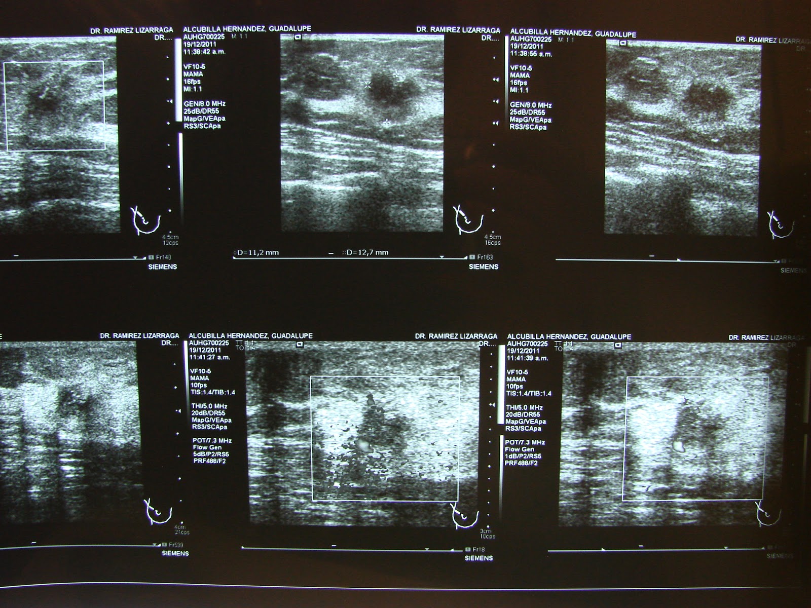
 Complementary Ultrasound IMage of the Left Lesion :
(remember after Biopsy) 


Morphology and Dimensions defined , anatomical concepts EASILY understood for the common health provider.....


I hope you agree with me as an Oncologist , enough suspicion is observed in order to recommend an open Biopsy of the LEFT nodule and in my opinion a radiologicaly guided on the RIGHT.

NOPE that did not happened , she was only sent for HER clinically evident left lesion.

So here I am with my Infrared Detector and stablishing or Aiding my Second Oncological Opinion :

Digital Infrared Analysis as a Complementary procedure in my patient:

 Basal Anterior View , infrared radiation is more obvious ont the left side , Mammary folds Irregularities , Left Axillary Predominance and an Isolated Thermal Summit coincidental with the Left PALPABLE lesion. 

But on the Right Side in the UPPER OUTER QUADRANT there is a Single Hyperthermia , that clinically is coincidental with the microcalcifications found on the Xray but not classified as SUSPICIOUS ( in the radiologist point of view) 


Physiologic series after cold challenge the same Hyperthermia is demostrated again.

Comment : Palpable Lesions are easy to biopsya ,and indications for them to be Biopsied are clear.

Infrared can Help :

"IDENTIFY SUBCLINICAL PALPABLE LESIONS"

In expert Hands (Oncologists Mainly) as a second look or opinion procedure is SAFE and can HELP Radiologists defined more clearly what they "consider" normal or abnormal.

Patient is on her way for BILATERAL BIOPSY ..results to be announced in the following weeks.

Keep posted and " open your minds for  a better future." EMC

Have a great weekend. 

lunes, 22 de abril de 2013

Search for , find and refer.


Digital Infrared Analysis of a NON PALPABLE Breast Invasive Ductal Carcinoma 1.2 cm.

Mammography is our cornerstone in detection , is it? Too complex strategy for most countries and their women. 

Sadly , Seems to me is not enough .......we should add valuable information and cases like this to flight the common enemy : Breast Cancer.

If you look for :


  1. Assimetry 
  2. Increased Vascular network
  3. Mammary fold and axillary heat differences.
Then you could help find with a little budget this specific PATIENTS with breast cancer  and Have a prompt and fast referal.

"To revolutionize the current System , ideas and facts can make a difference." EMC.

Have a great week.

lunes, 15 de abril de 2013

Hallo Deutschland

Grüße zu den deutschen Kollegebesuchern dieses Blogs. (Beispiele von den Kästen positiv für Brustkrebs mit der Diagnose schwierig oder durch technisches herkömmliches verzögert)

nwo es Diagnosezweifel gibt und voraussagt, dass es die Möglichkeit des Verbesserns ........ von BII weniger als 5% gibt, BIRADS III weniger als 10% und BIRADS IV das irreführendste von allen von 5 zu 95%) 

The\n\nfachkundige einzelne Interpretation ist 100% Abhängiges auf der Subjektivität anderer Radiologen

Hallo Deutschland: eine Entschuldigung für die möglichen Rechtschreibfehler:\n

An den letzten Tagen habe ich mit großer Freude bemerkt, dass die Hauptquelle von Besuchen zu diesem Blog von den deutschen Kollegen kommt und zum Eintritt oder zur Veröffentlichung mit der höchsten Anzahl von Schlägen das BIRADS III bezogen ist.\nIntelligenter Gesichtspunkt, verbessern die Qualität des BIRADS III.

Obgleich ich persönlich denke, dass das BIRADS IV ziemlich unspezifisch ist, denken Sie?\nJede mögliche Weise danken Ihnen für betroffen sein und ich bin an Ihrem Service, zum irgendwelcher Fragen oder Interessen von Mexiko Enrique in naher Zukunft zu lösen… ….
 Die Infrarotanalyse ermittelt, definiert und bestimmt die Sendungsbereiche quantitativ.

Die"metabolische" Ergänzung von dieser Art von vergleichbaren Informationen mit der kontralateralen Seite oder der anderen Drüse, kann die Qualität unserer Diagnose drastisch verbessern.

Guter Tag.


jueves, 4 de abril de 2013

Geographical And Anatomical Monitoring............

Every single diagnostic procedure or imaging technique as a matter of fact is an interpretation of any Physical or Chemical Process in the Human Body , in the vast majority of these :

The results they turn out represent a SINGLE moment in time....

High Blood Pressure is Confirmed after several times it is taken in different environments...

Diabetes or Glucose intolerance is better diagnoses after the serial measurements after stimuli or with a "curve" definition.

Arrythmia has a more clear diagnosis when a "Holter" : 24 hour monitoring is done.

In breast cancer this MONITORING system has been historically determined to be every 365 to 730  days after 40 or 50 years of age.

Because MORPHOLOGIC changes take time to APPEAR in our Current Gold Standard Methods : Mammo , Ultrasound ,Nuclear Medicine or MRI.

Here we are safe and evident ......right?  Are we?

Yet around 50% of the cases are diagnosed before 50 years of age.
70% of the cases are palpable and even will large scale screening : at best (developed countries )  around 30% are diagnosed at a non palpable stage.

Not to mention overdiagnosis , overtreament and "benign" natural evolution of certain in situ forms of the disease.

What if we could insert a closer in time monitoring system for breast cancer , especially the High Grade or aggresive forms of the disease?

What if we could combine different informations from different Physical Interpretation Angles.

Here is  a resemblance that dignifies this possibility:


Anticipating an eruption
It is not yet possible to predict volcanic eruptions, but several new monitoring techniques, which are being tried out at Stromboli and elsewhere, may allow scientists to improve early warning of eruptions.

In many volcanoes, the bubbles of steam that drive the eruptions are also rich in carbon dioxide, sulphur dioxide and hydrogen chloride. These gases can be detected using chemical sensors, and spectrometers (both hand-held and on satellites), and changes in the proportions of the different gases leaking out of the volcano are often a sign of the arrival of new magma at depth. In some volcanoes, the slow rise of magma towards the surface leads to pressure changes inside the volcano, causing it to swell up. This swelling 
can be measured by radar instruments on orbiting 
satellites, and these signals are increasingly begin used 
to monitor the behavior of restless volcanoes. 

Volcanoes themselves are also noisy, with small earthquakes triggered by the breaking open of new fractures as magma rises. The bubbly melt trapped within the plumbing system of the volcano can also act as a sound-box, leading to distinctive signals as the seismic waves from local earthquakes resonate inside the volcano.


Closer monitoring systems that combine useful information ,

 adjusted in time in a accurate format with an 

understandable meaning can predict future events with no 

doubt at all.

Although historically mentioned by some , spectrometry or 

infrared or thermal monitoring of the breast 


In Vivo..............sounds likely  and highly possible ....


Would you like to be involved in a project like this?


Now here is an Infrared Image of a NON palpable aggresive 
invasive Breast Carcinoma in a High Risk Patient.


Surveillance , monitoring  of any type includes comparative images between "healthy" or Silent vs "diseased" or prompt to erupt.....


Infrared Close up of the Left "Diseased" Breast  or "Erupting" Volcano.

What if she had previous access to a Metabolic Breast Monitor?

Would you like to see the XRay images ? Keep Posted Please.....


lunes, 18 de marzo de 2013

Glimpse to the future .


FACTS: 
  • Thermography historically has failed as an screening procedure.
  • Ultrasound is not recommended as viable option too
  • MRI is unavailable and quite expensive for it to be an standard all population method.
  • And Mammography our champion is currently in the edge of criticism mainly because of its cost benefit ratio -overdiagnosis + overtreatment numbers and its consequences.

YET : 

They all HAVE a main disadvantage : 

  1. TIME and INTERVAL ( Initial screening procedure, frequency)
AND:

IN MOST CASES IT COULD BE A YEAR OR MORE.....


Now:

What if there could be a method of 

CONTINUOS  MONITORING......... 

That is right the only logical option is thermal or infrared monitoring.

I believe with our current knowledge and technology , Thermometry in any of its forms , coupled to current standard recommendations and methods  , clinical or radiological , will eventually assist detection of breast cancers, Specially the high metabolic ones or the aggresive forms of the disease.

Here is an example...... hope you enjoy it.......


Remember the last case : BIRADS III Left breast that eventually turned out to be a breast cancer?

And then 2 years later a secondary CONTRALATERAL breast cancer detected or pin pointed by IR image previously?


Clinical "drawing" of the original 1 cm nodule , and the corresponding infrared image.
In some cases and locations , some tumors might not be detected by mamogram either because of density , technical or anatomical reasons.


DIRA interpretarion demostrated a higher score for the left breast , but also pin pointed independently high infrared emission areas on the right breast.....


Now 2 years later:

Infrared Images after treatment , clearly signal RIGHT area , now well defined as the Highest IR peak or "Thermal Summit" 


IR as I mentioned earlier  could assist , enhance and improves  performance of  Breast Physical Exam and could guide Ultrasound for millimetrical or close to 1 cm lesions.....NOT BAD i Think.


Here comes a vision..........already mentioned in 2007 trying to introduce the first "Smart Bra" , with the fact that even 6 years later nothing has happened.

And in last October a company in the US published their own system with great expectation for the future.

So : what if my patient would have been wearing such a device or a monitoring system:


No Comments , except that : 

The images are clear enough for anyone , the message is simple , and evidence of that new possibility is actually in the right path.....

"Intelligent Thermal or Infrared Monitoring systems WILL aid detection of breast cancers and can be safely introduced in the Clinical Care for our Patients."EMC

I have no other option but to commit myself into this idea and mission.

The How , Who and When is up to you and your collaboration.............

Have a nice day.


lunes, 25 de febrero de 2013

"Breast Thermography can predict and detect 8-10 years before than a mammogram"

Breast Thermography SUPPORTERS STATE  that:

 IR can detect changes before cancer is actually seen..................

I personally and certainly don´t think so it is very hard to believe  ( Not in our science and evidenced based world , not with previous technology or historical facts ) 

Here comes my and probably my colleagues question to this statement  :

"So , You tell me now but you prove it 8 to 10 years later? "

or 

"How can I perform a Biopsy in an absent to Ultrasound , MRI or Mammogram   Non Morphological Infrared Change ?"

SHAKY GROUNDS INDEED.

But ; regardless of what I thought or knew ...........time and patience brought to me  a case that can clearly support at least that this is actually not only possible but opens the door to RETHINK and REEXPLORE........


Female patient asymptomatic in her early forties until she felt a non painful LEFT breast nodule . BIRADS III you agree and surveiled under standard of care 6 months later , going from 5 to 1.1 cm in size 


After cold stress , the highest metabolic area in the left breast is highlightened clearly. 


 Digital Infrared Analysis , PINPOINTS the nodule (Arrow) 

 She underwent lumpectomy and Sentinel Lymph Node disection , Chemotherapy Radiotherapy and Tamoxifen as Hormotherapy , NOW : 
In follow consultation  , after feeling an irregular superior RIGHT area  I Performed DIRA and I asked for radiological mammo and ultrasound

 Self explaining 

So 2012 now present with a MORPHOLOGICAL visual change that was pinpointed or Insinuated since 2010

SERENDIPITY? 
Maybe..........................But Maybe Not 
Keep posted so we can discuss pathological findings.


miércoles, 13 de febrero de 2013

BIRADS III , To Biopsy or not to Biopsy!

Great week you all ..!
Last night I was having a look at my Blog's statistics.
And I reached the conclusion that the sample case with more hits was:

BIRADS III and Infrared......

So I must think that although you do not comment at all (who would , this issue is too controversial even to speak about it....)
I sense that the majority of my readers should be breast cancer specialists ( radiologists , oncologists, Gynecologists or breast surgeons)

If I am right you can have a smile at home!!!!

Well keep in touch........BIRADS III , to BiRopsy or not to BiRopsy. ( Visit and or Join The BIRADS/Infrared Project)
at:
A promise is a debt : 
A few year ago a 40 years of age asymptomatic  patient of mine underwent an screening mammogram and  handled this images:


Medio Lateral Oblique view , evident density assimetry in the upper outer right quadrant.


Close up : No Microcalcifications are seen , yet I might agree retrospectively you can argue architectural distorsion and irregular profile. BIRADS 0 went to a complementary Ultrasound: 


An heterogeneous Morphological Lesion is found , with cystic and solid components almost circular in its form no posterior "shadow" is seen .

FINAL RADIOLOGIC DIAGNOSE BIRADS III , close follow up right? 

I WOULD THINK SOME MAY ARGUE AN ERRONEOUS OR MISTAKEN DIAGNOSIS , YET IT HAPPENED. 

Then she attended 3 months later to a SECOND Opinion.

So this are her DIRA images:


Basal DIRA series , anterior and oblique views , upfront as any NEW language or Unknown one there should be a little doubt an distrust.

If you look up closely actually there is a little assimetry that depends on the upper outer quadrant of the Right Breast what we might call  THE INTEREST AREA or IA.


After cold challenge the Functional or Physiological series were taken. No obvious difference for the UNTRAINED eye. 


Yet with the Software "Isotherm" application a Slight difference is then seen in comparison to the left side (ARROW) it actually is coincidental to the  physical finding of an "irregularity" zone at palpation . 


Same image but different color option.

So now I am thinking :" there is a HIGH METABOLIC area that is coincidental with my MORPHOLOGICAL findings by standard mammogram and ultrasound. Not to mention that this area by incidence is the highest for a Breast Cancer to appear." EMC

Recommendation : I think you should perform not in 6 months but
 as soon as possible a Mammography  and corresponding ultrasound. 

These are the images taken :


Cefalocaudal view , density assimetry now is evident.


Oblique view , confirm previous findings.


Close up : Highly suspicious Microcalcifications are NOW EVIDENCED


Corresponding Ultrasound Images : NOW MORPHOLOGICAL CLEAR CHANGES ARE SEEN AND DOPPLER DEMONSTRATES INCREASED VASCULAR FLOW.

BIRADS V .....

Most would argue that based on experience diagnosis could have been different previously. Better yet if there is a Close Surveillance the same diagnostic algorithm would have been done. And even if DIRA proofs a benefit in helping DIAGNOSIS and DETECTION , prognosis would be the same.

Reality : BIRADS III is based on INDIVIDUAL RADIOLOGIST Criteria , Follow up sometimes cannot be achieved. 
And BIRADS II was another possibility for the first classification.

The ecuation is simple:
"BIRADS III (even IVa) Morphologically proven , DIRA coincidental for HIGH METABOLIC ACTIVITY Plus ONCOLOGICAL experience : Recommend for SURE to Biopsy."EMC

These are the images taken in the OR , a possible next level.
Keep in touch.............


The idea is to gather BIRADS III lesions internationally , I will creat this next blog for those who would like to participate......
But now I am going to celebrate my Boy´s Birthday so I guess we will see you around next week.

http://birads3infraredproject.blogspot.mx/