Just Now I received an interesting clinical case , which I would Like to SHARE:
- FEMALE
- 45 years of age.
- First Birth at 17
- Positive Breast Feeding
- 8 Years with Oral Contraception
- Last Mammography 18 months.(REPORTED AS NORMAL BIRADS II )
- I week of Left Mastalgia and other minimal symptoms .
- Non identified Palpable Lesion on either Breast
- Axilla : Undetermined 1 cm palpable Lymph Node on the left side.
REMEMBER : "No radiological exams at all with the following Infrared Images" :
I will talk you through them:
Basal Anterior View :
Obvious Left Assimetry
Enhanced Left Vascular Network
Irregular Left Mammary Fold
Single Upper Quadrant (12hrs) Line B on the Left Side.
Slightly Retracted Left Nipple....
Non of the previous findings on the Right...
Oblique View of the Left Breast , notice how entangled is the vascular network....
Physiological or Post Cold Stress Challenge View:
Normal Right Breast cools down
Abnormal metabolic and Vascular Left Changes are enlightened.
Single Hiperthermia on the Left.
INFRARED ANALYSIS HARBORS :
Normal Right Breast cools down
Abnormal metabolic and Vascular Left Changes are enlightened.
Single Hiperthermia on the Left.
INFRARED ANALYSIS HARBORS :
- POSITIVE and NEGATIVE PREDICTIVE VALUES FOR DETECTED LESION
- METABOLIC BEHAVIOUR OF A DETECTED LESIONS
- CAN STABLISH A RISK FOR FUTURE CANCER SPECIALLY IN FAMILIAL CASES AS THIS PATIENT IS.
- CAN MONITOR EACH BREAST AS AN INTERVAL PROCEDURE.
"For me there is no doubt at all , Digital Infrared Analysis of the Breast holds an evident truth to be rediscovered by Breast specialists , hopefuly radiologists and Oncologists." EMC.
BIRADS III PLUS A POSITIVE METABOLICAL EXAMINATION SHULD PROMPT THE NEED FOR BIOPSY.
(And you can quote me on that)
BIRADS III PLUS A POSITIVE METABOLICAL EXAMINATION SHULD PROMPT THE NEED FOR BIOPSY.
(And you can quote me on that)
Keep posted : ANATOMICAL OR MORPHOLOGICAL IMAGES will arrive soon.
My prediction : Positive Case for Breast Carcinoma , Invasive i would say.
"Prediction is often confused with witchery , specially when the language is UNKNOWN......"
Lets see what happens...................
June 3rd 2013 .
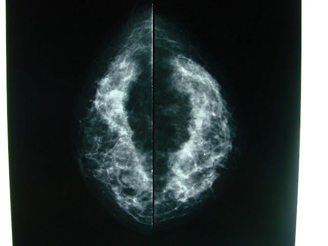
Patient arrived with the following Mammographical and Ultrasound Images:
Just to point out credibility , done last week and 45 years of age .........
Close Up............. density becomes more evident right?
Cefalo Caudal Views , subtle central and inner density assimetry in the left breast with the same calcification , a little loss of the retromammary space
I WILL QUOTE : " Left breast with a higher echogenicity , diffuse , with some microcysts and ductal dilations ,
Elastography showed : left associated mixed regions some with a higher or more "solid" nature....
June 3rd 2013 .
Patient arrived with the following Mammographical and Ultrasound Images:
Standard Oblique views , there is a density assimetry in the left breast with a single possibly benign calcification , no architectural distorsion or spiculated lesion. Inflammatory axillary lymph Nodes.
Close Up............. density becomes more evident right?
Calls my attention a single Level 1 Lymph node , which has higher density in it´s hilium......makes me wonder now....
Complementary Ultrasound , plus doppler :
Elastography showed : left associated mixed regions some with a higher or more "solid" nature....
Now here comes UNCERTAINTY : BIRADS III
Standards of care demand a 6 month follow up
Lets BEND the rules , since Clinical knowledge comes from the sum of all previous cases...
I recommended no matter what happens after antiinflammatory treatment and antibiotic to perfom CBC , Coagulation , and glucose tests.
And after reviewing the results , i will schedule a Biopsy with a trucut.....
OBVIOUS 2 POSSIBILITIES:
MASTITIS vs Breast Carcinoma possibly T4d : inflammatory.
What do you THINK ? .............well:
"Infrared images are intended ONLY for Breasts Specialists , Breast Cancer Treatment is responsability ONLY for Oncologists.
Detection , Diagnosis and Treatment action depends entirely from : Evidence , History , Experience and a "different sense of feeling" only reserved for those Real Clinitians..........EMC"
(the ones that do not rely only in what they were told , those who challenge with patience and good intention.... )


No hay comentarios:
Publicar un comentario