Breast Digital Infrared Analysis (DIRA) , a forgotten and set aside procedure.

Visita: termografiamamaria.blogspot.com (Version en Español)

Breast DIRA (previously named Thermography) already proved in the past that it´s use as an screening procedure had low specificity and high sensitive values for it to be recommended as an standard care Breast diagnosis tool .
Yet , until recently (last 5 years) active research mainly by Surgical Oncologists or Breast Surgeons have published interesting results.( Cornell NY , Addenbrooke Cambridge , Ville Marie Montreal , Taiwan and Mexico CEPREC)
FDA previous published criteria were prohibiting DIRA as well as the A.C. of Radiologists ( as an initial SCREENING TOOL)

Yet it has been cleared by the FDA approval as an adjunctive screening tool for breast cancer .( under 510 k) .

DIRA´s meaning is not an ANATOMIC or ARCHITECTURAL procedure ,it is a METABOLIC or PHYSIOLOGIC ONE so at first it ´s current recommendation should be only as a COMPLEMENTARY STUDY.

As such DIRA could help in specific scenarios, BIRADS 0,3,4 and give potential useful additional data in BIRADS5. EVEN BEFORE BIOPSY
Could aid mammogram in young,dense and breast prosthesis patients, and should be evaluated in other clinical scenarios as a Neoadyuvant Monitoring tool.
Finally as an early detecion procedure specifically in underdeveloped countries , could Triage in search of aggresive forms of the disease(T1,G2_G3). And aid Physical Exam of the Breast .
This site is dedicated to open mind Breast specialists ONLY .
¨EXPANDING OUR CURRENT COMPREHENSION, UNDERSTANDING AND APPROACH OF IMAGE AND DIAGNOSIS IN BREAST CANCER¨

lunes, 24 de junio de 2013

Digital Infrared Analysis of the Breast_Free and Available.

Breast Cancer ORDER is mantained by the evidence created during the last 40 years with Mammography.

Nobody denies its intrinsic value and strengths.

It is our primary language against the Common Enemy : Breast Cancer

Yet , to reach its ideal goals and current trends it involves a complicated network of :


  1. personal , 
  2. political , 
  3. cultural , 
  4. geographical and 
  5. economical interests around this sometimes regretfuly "fashion" topic.


(Specially sensitive and a super candidate for promotion and glamour.)

To affirm that ONLY mammography holds the truth is 

To deny that anything else can help , and sounds to me as a MONOPOLY ...........

A scientifically based evidence of the INFRARED analysis and Behaviour of those MALIGNANT potentially MORTAL types of BREAST CANCER  in my mind is not only necessary but an obligation.....for all 

Since current trends will  be of Tsunamic Proportions as some have already stated 

.....Advanced cases in the vast majority of countries will keep on rising if we follow the same recommendations.

I do not intend to set aside Mammography ...that is not only impossible but Stupid.

I simply would like to let you know that DIRA can be useful and as time goes by , more and more examples will join , more women will be helped and an specific nich for its use will rise.

Last of all : I think that DIRA should be FREE  or near to free and available only for Breast Cancer Specialists .

Have a great week------EMC

jueves, 20 de junio de 2013

Nanoparticles-Infrared Image-Excised MArgins in Breast Cancer.

Hi everybody , one of the most important problems that we as surgeons have in breast conserving surgery is being able to deliver : SAFE MARGINS.

This is generally achieved as a "BLIND" procedure and it is dependent of individual  clinical and surgical  expertise  in order to produce negative tissue inked margins in the final pathological report , some recommend at least 1 cm of safe distance... so 

QUESTION : Can we in the near Future have an available method that can help us assure this "safety" in margins ?

Recent efforts are being described that couple:


  1.  Nanoparticles associated with 
  2. CANCER SPECIFIC Antibodies 
  3. Nanoparticle excited physical properties  and  
  4. It corresponding image :
Here is an example and is reference ......

I believe : 

"In the near future we will have the Technology by the association of Nanoparticles , monoclonal antibodies and the digital infrared image or another physical and visual intrinsic characteristic , which : 

WILL OFFER in vivo A VISUAL AID FOR THE SURGEON to produce safe margins in breast  conserving surgery ."EMC

Have a great day : 


(A) Schematic representation of nanoparticle communication to achieve amplified tumor targeting. Tumor-targeted signaling nanoparticles (blue) broadcast the tumor location to the receiving nanoparticles (red) present in circulation. (B) Shown are the harnessing of the biological cascade to transmit and amplify nanoparticle communication and the molecular signaling pathway between the signaling and receiving components. (C) Thermographic images of the photothermal NRs with heating. Seventy-two hours after NR or saline injection, mice were co-injected with FXIII-NWs and untargeted control-NWs, and their right flanks were broadly irradiated (top). Twenty-four hours post-irradiation, whole-animal fluorescence imaging revealed the distribution of the receiving nanoparticles (bottom). (D) Amplified tumor therapy with communicating nanoparticles. Tumor volumes following a single treatment with the communicating nanoparticle systems and controls. Reproduced with permission from ref :

 von Maltzahn G, Park JH, Lin KY, Singh N, Schwöppe C, Mesters R. et alNanoparticles that communicate in vivo to amplify tumour targetingNat Mater. 2011;10:545-52


This is another example with Fluorescent propierties...... 

Assessing Breast Cancer Margins Ex Vivo Using Aqueous Quantum-Dot-Molecular Probes
International Journal of Surgical Oncology
Volume 2012 (2012), Article ID 861257, 12 pages
doi:10.1155/2012/861257

miércoles, 29 de mayo de 2013

BIRADS III THERE SHOULD BE NO DOUBT AT ALL .....

Hi again :

Just Now I received an interesting clinical case , which I would Like to SHARE:

  1. FEMALE
  2. 45 years of age.
  3. First Birth at 17
  4. Positive Breast Feeding
  5. 8 Years with Oral Contraception
  6. Last Mammography 18 months.(REPORTED AS NORMAL BIRADS II ) 
  7. I week of Left Mastalgia and other minimal symptoms .
  8. Non identified Palpable Lesion on either Breast 
  9. Axilla : Undetermined 1 cm palpable Lymph Node on the left side. 

REMEMBER : "No radiological exams at all with the following Infrared Images" : 

 I will talk you through them: 


Basal Anterior View : 
Obvious Left Assimetry 
Enhanced Left Vascular Network
Irregular Left Mammary Fold
Single Upper Quadrant (12hrs) Line B on the Left Side.
Slightly Retracted Left Nipple....


Non of the previous findings on the Right...


Oblique View of the Left Breast , notice how entangled is the vascular network....


Physiological or Post  Cold Stress Challenge View:
Normal Right Breast cools down
Abnormal metabolic and Vascular Left Changes are enlightened.
Single Hiperthermia on the Left.

INFRARED ANALYSIS HARBORS :

  1. POSITIVE and NEGATIVE PREDICTIVE VALUES FOR DETECTED LESION
  2. METABOLIC BEHAVIOUR OF A  DETECTED LESIONS
  3. CAN STABLISH A RISK FOR FUTURE CANCER SPECIALLY IN FAMILIAL CASES AS THIS PATIENT IS. 
  4. CAN MONITOR EACH BREAST AS AN INTERVAL PROCEDURE.
"For me there is no doubt at all , Digital Infrared Analysis of the Breast holds an evident truth to be rediscovered by Breast specialists , hopefuly radiologists and Oncologists." EMC.

BIRADS III PLUS A POSITIVE METABOLICAL EXAMINATION SHULD PROMPT THE NEED FOR BIOPSY.

(And you can quote me on that)

Keep posted : ANATOMICAL OR MORPHOLOGICAL IMAGES will arrive soon.

My prediction : Positive Case for Breast Carcinoma , Invasive i would say.

"Prediction is often  confused with witchery , specially when the language is UNKNOWN......"

Lets see what happens...................

June 3rd 2013 .
Patient arrived with the following Mammographical and Ultrasound Images:

 Just to point out credibility , done last week and  45 years of age .........

Standard Oblique views , there is a density assimetry in the left breast with a single possibly benign calcification , no architectural distorsion or spiculated lesion.  Inflammatory axillary lymph Nodes. 


Close Up............. density becomes more evident right?


Calls my attention a single Level 1 Lymph node , which has higher   density in  it´s hilium......makes me wonder now....


 Cefalo Caudal Views , subtle central and inner density assimetry in the left breast with the same calcification , a little loss of the retromammary space 

Complementary Ultrasound , plus doppler : 

 I WILL QUOTE : " Left breast with a higher echogenicity , diffuse , with some microcysts and ductal dilations , 
 Elastography showed : left associated  mixed regions  some with a higher or more "solid" nature....

Now here comes UNCERTAINTY : BIRADS III

Standards of care demand a 6 month follow up

Lets BEND the rules , since Clinical knowledge comes from the sum of all previous cases...

I recommended no matter what happens after antiinflammatory treatment and antibiotic  to perfom CBC , Coagulation , and glucose tests. 

And after reviewing the results , i will schedule a Biopsy with a trucut.....

OBVIOUS 2 POSSIBILITIES:

MASTITIS vs Breast Carcinoma possibly T4d : inflammatory.

What do you THINK ? .............well:

"Infrared images are intended ONLY for Breasts Specialists , Breast Cancer Treatment is responsability ONLY for Oncologists.
Detection , Diagnosis and Treatment action depends entirely from : Evidence , History , Experience and a "different sense of feeling" only reserved for those Real Clinitians..........EMC"

(the ones that do not rely only in what they were told , those who  challenge with patience and good intention.... ) 


jueves, 23 de mayo de 2013

Is an Infrared Analysis of the Breast a complementary procedure?

SIMPLE :

If an Infrared Analysis of the Breast is an approved  Complementary procedure , then why don't we use it as SUCH?

ANSWER : NOT A SINGLE RADIOLOGIST ( except in Brasil ) has been trained or  INTERESTED in it  and as consequence the vast majority of Oncologists worldwide........


THE TRUTH : There are some BIRADS III that eventually turn into a positive cancer cases

                         BIRADS IV is way TOO tricky and HOLDS LOW SPECIFICITY.

                         And BIRADS 0 obviously needs SOME help.


Infrared Analysis HOLDS a physical and Biological MEANING , NEW evidence is  waiting to be built by EXPERTS in Breast Diseases , not in "alternative" health providers.

Seems logical to me and to you?


lunes, 13 de mayo de 2013

Is Mammography an Stereotyped Procedure?

Good Morning , I woke up this morning with this question:

Is mammography an stereotype behavior or medical , radiological procedure?

I was taught that it is our better weapon against Breast Cancer.
I was taught that chances of surviving depend ONLY in is detection Capabilities.
I was taught it should be done annually starting age at 40 .
I was taught it lowered by 30 % mortality rates.
I was taught never to question or challenge this concepts.

BUT

Is it really our best weapon when IT SAVES In situ lesions never to develop clinical signs or even influence mortality?
Not to mention over diagnosis and over treatment ....
Does surviving breast cancer Excludes then our current BETTER Treatments?
What about women 40 years and younger? And how about evidence against Annual vs BIANNUAL?
And  starting until 50 (normal risk)
Was it really 30% reduction in mortality or closer to 10%?

I think we Should we Challenge stereotypes and prove them wrong (when they are ) , and accept ADJUNCTIVELY WITH THE SATNDARDS OF CARE  some "old"  but 

  • logical , 
  • physical , 
  • reproductible , 
  • and methodical with a possible use , for a Better Detection , Less false positive Biopsies and help current recommended procedures.


Stereotype means : "SOLID IMPRESSION"

I believe we are entitled to rearrange strategies based on facts and evidenced associated to specific real human environments and scenarios....

" Not everything that shines is gold, is it? " EMC

Good day.

lunes, 29 de abril de 2013

Raise your hand?


Positive Breast Cancer all of them 

NON PALPABLE :


It May sound ridiculous , but some IR specific patterns actually mean :

INVASIVE BREAST CANCER...........

These could Help  : 

DETECTION . DIAGNOSIS or TREATMENT and even FOLLOW UP-

Keep it up.....it really does not matters at all how you identify a Breast Cancer if you actually can , right?

I Challenge this little evidence to be proved or revoked..........who will raise his or her hand?


miércoles, 24 de abril de 2013

Availability , prompt attention and Specialized Diagnostic Aid.

Hi , with regret the following Surgical Oncology Scenario is quite common : 

Female on her early 50´s no previous history for breast cancer in her family , no other risk factors. 

  1. Palpable lesion on the left breast : NON CONCLUSIVE post OPEN biopsy pathological diagnosis and persistance of the original tumor near to the scar and undeterminate axillary lymph nodes
  2. Refered after a 2nd  NEGATIVE trucut biopsy to SURGICAL complete removal.

Started seeking for medical attention 13 , yes 13 Months previously with the following standard images:

(Remember please , unfortunately availability of excellent breast cancer diagnosis is far from ideal in the vast majority of  countries , in mine (Mexico)  it is estimated that after radiological suspicion for open population detection  , women start treament at least 9 months later.................some thing to think about and work for i believe  ) :(

Here they are:


 Dense Breast Tissue ++++
 Some Microcalcifications are seen Upper and 
 Outer Quadrant on the RIGHT side (remember palpable nodule on the LEFT) 
 Close up and amplification view: 


 Complementary Ultrasound IMage of the Left Lesion :
(remember after Biopsy) 


Morphology and Dimensions defined , anatomical concepts EASILY understood for the common health provider.....


I hope you agree with me as an Oncologist , enough suspicion is observed in order to recommend an open Biopsy of the LEFT nodule and in my opinion a radiologicaly guided on the RIGHT.

NOPE that did not happened , she was only sent for HER clinically evident left lesion.

So here I am with my Infrared Detector and stablishing or Aiding my Second Oncological Opinion :

Digital Infrared Analysis as a Complementary procedure in my patient:

 Basal Anterior View , infrared radiation is more obvious ont the left side , Mammary folds Irregularities , Left Axillary Predominance and an Isolated Thermal Summit coincidental with the Left PALPABLE lesion. 

But on the Right Side in the UPPER OUTER QUADRANT there is a Single Hyperthermia , that clinically is coincidental with the microcalcifications found on the Xray but not classified as SUSPICIOUS ( in the radiologist point of view) 


Physiologic series after cold challenge the same Hyperthermia is demostrated again.

Comment : Palpable Lesions are easy to biopsya ,and indications for them to be Biopsied are clear.

Infrared can Help :

"IDENTIFY SUBCLINICAL PALPABLE LESIONS"

In expert Hands (Oncologists Mainly) as a second look or opinion procedure is SAFE and can HELP Radiologists defined more clearly what they "consider" normal or abnormal.

Patient is on her way for BILATERAL BIOPSY ..results to be announced in the following weeks.

Keep posted and " open your minds for  a better future." EMC

Have a great weekend. 

lunes, 22 de abril de 2013

Search for , find and refer.


Digital Infrared Analysis of a NON PALPABLE Breast Invasive Ductal Carcinoma 1.2 cm.

Mammography is our cornerstone in detection , is it? Too complex strategy for most countries and their women. 

Sadly , Seems to me is not enough .......we should add valuable information and cases like this to flight the common enemy : Breast Cancer.

If you look for :


  1. Assimetry 
  2. Increased Vascular network
  3. Mammary fold and axillary heat differences.
Then you could help find with a little budget this specific PATIENTS with breast cancer  and Have a prompt and fast referal.

"To revolutionize the current System , ideas and facts can make a difference." EMC.

Have a great week.

lunes, 15 de abril de 2013

Hallo Deutschland

Grüße zu den deutschen Kollegebesuchern dieses Blogs. (Beispiele von den Kästen positiv für Brustkrebs mit der Diagnose schwierig oder durch technisches herkömmliches verzögert)

nwo es Diagnosezweifel gibt und voraussagt, dass es die Möglichkeit des Verbesserns ........ von BII weniger als 5% gibt, BIRADS III weniger als 10% und BIRADS IV das irreführendste von allen von 5 zu 95%) 

The\n\nfachkundige einzelne Interpretation ist 100% Abhängiges auf der Subjektivität anderer Radiologen

Hallo Deutschland: eine Entschuldigung für die möglichen Rechtschreibfehler:\n

An den letzten Tagen habe ich mit großer Freude bemerkt, dass die Hauptquelle von Besuchen zu diesem Blog von den deutschen Kollegen kommt und zum Eintritt oder zur Veröffentlichung mit der höchsten Anzahl von Schlägen das BIRADS III bezogen ist.\nIntelligenter Gesichtspunkt, verbessern die Qualität des BIRADS III.

Obgleich ich persönlich denke, dass das BIRADS IV ziemlich unspezifisch ist, denken Sie?\nJede mögliche Weise danken Ihnen für betroffen sein und ich bin an Ihrem Service, zum irgendwelcher Fragen oder Interessen von Mexiko Enrique in naher Zukunft zu lösen… ….
 Die Infrarotanalyse ermittelt, definiert und bestimmt die Sendungsbereiche quantitativ.

Die"metabolische" Ergänzung von dieser Art von vergleichbaren Informationen mit der kontralateralen Seite oder der anderen Drüse, kann die Qualität unserer Diagnose drastisch verbessern.

Guter Tag.


jueves, 4 de abril de 2013

Geographical And Anatomical Monitoring............

Every single diagnostic procedure or imaging technique as a matter of fact is an interpretation of any Physical or Chemical Process in the Human Body , in the vast majority of these :

The results they turn out represent a SINGLE moment in time....

High Blood Pressure is Confirmed after several times it is taken in different environments...

Diabetes or Glucose intolerance is better diagnoses after the serial measurements after stimuli or with a "curve" definition.

Arrythmia has a more clear diagnosis when a "Holter" : 24 hour monitoring is done.

In breast cancer this MONITORING system has been historically determined to be every 365 to 730  days after 40 or 50 years of age.

Because MORPHOLOGIC changes take time to APPEAR in our Current Gold Standard Methods : Mammo , Ultrasound ,Nuclear Medicine or MRI.

Here we are safe and evident ......right?  Are we?

Yet around 50% of the cases are diagnosed before 50 years of age.
70% of the cases are palpable and even will large scale screening : at best (developed countries )  around 30% are diagnosed at a non palpable stage.

Not to mention overdiagnosis , overtreament and "benign" natural evolution of certain in situ forms of the disease.

What if we could insert a closer in time monitoring system for breast cancer , especially the High Grade or aggresive forms of the disease?

What if we could combine different informations from different Physical Interpretation Angles.

Here is  a resemblance that dignifies this possibility:


Anticipating an eruption
It is not yet possible to predict volcanic eruptions, but several new monitoring techniques, which are being tried out at Stromboli and elsewhere, may allow scientists to improve early warning of eruptions.

In many volcanoes, the bubbles of steam that drive the eruptions are also rich in carbon dioxide, sulphur dioxide and hydrogen chloride. These gases can be detected using chemical sensors, and spectrometers (both hand-held and on satellites), and changes in the proportions of the different gases leaking out of the volcano are often a sign of the arrival of new magma at depth. In some volcanoes, the slow rise of magma towards the surface leads to pressure changes inside the volcano, causing it to swell up. This swelling 
can be measured by radar instruments on orbiting 
satellites, and these signals are increasingly begin used 
to monitor the behavior of restless volcanoes. 

Volcanoes themselves are also noisy, with small earthquakes triggered by the breaking open of new fractures as magma rises. The bubbly melt trapped within the plumbing system of the volcano can also act as a sound-box, leading to distinctive signals as the seismic waves from local earthquakes resonate inside the volcano.


Closer monitoring systems that combine useful information ,

 adjusted in time in a accurate format with an 

understandable meaning can predict future events with no 

doubt at all.

Although historically mentioned by some , spectrometry or 

infrared or thermal monitoring of the breast 


In Vivo..............sounds likely  and highly possible ....


Would you like to be involved in a project like this?


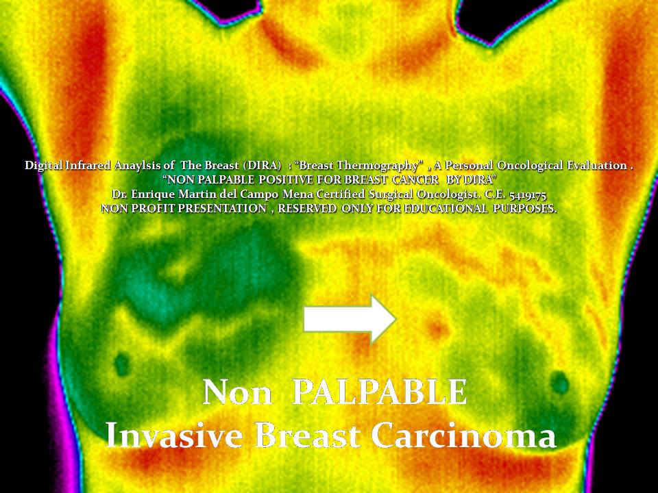
Now here is an Infrared Image of a NON palpable aggresive 
invasive Breast Carcinoma in a High Risk Patient.


Surveillance , monitoring  of any type includes comparative images between "healthy" or Silent vs "diseased" or prompt to erupt.....


Infrared Close up of the Left "Diseased" Breast  or "Erupting" Volcano.

What if she had previous access to a Metabolic Breast Monitor?

Would you like to see the XRay images ? Keep Posted Please.....